

Fluorescent-Labeled Antibodies for Western Blot: High-Sensitivity and Multiplex Detection Solutions
In the field of protein expression analysis, Western Blot remains one of the most commonly used and reliable foundational techniques. However, as research progresses from simply determining whether a protein is expressed to examining changes in expression levels, multi-target regulation, and signaling pathway analysis, traditional antibody systems based on chemiluminescent or colorimetric detection increasingly reveal limitations in quantitative accuracy, dynamic range, and multiplexing capability. The emergence of fluorescent-labeled antibodies provides a more stable, quantifiable, and multiplex-compatible approach for Western Blot detection, becoming an essential tool in modern protein research.
What Are Fluorescent-Labeled Antibodies?
Fluorescent-labeled antibodies are bioanalytical tools in which fluorescent dye molecules are stably attached to antibodies through chemical conjugation or biochemical modification. After binding to specific target proteins, these antibodies emit a fluorescent signal under a specific excitation wavelength, enabling protein localization, quantification, and multi-target analysis. Compared with traditional enzyme-labeled antibodies, fluorescent-labeled antibodies offer significant advantages in signal linearity, stability, and multi-channel detection capability, making them the preferred choice for Western Blot, high-throughput protein analysis, and multiplex immunoassays.
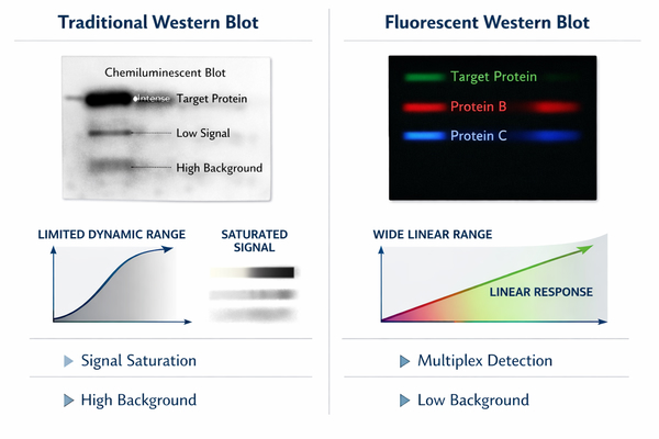 Fig. 1. Conventional vs fluorescent western blot (BOC Sciences Authorized).
Compared with traditional enzyme-labeled antibodies, fluorescent-labeled antibodies offer significant advantages in signal linearity, stability, and multi-channel detection capability, making them the preferred choice for Western Blot, high-throughput protein analysis, and multiplex immunoassays.
Why Use Fluorescent-Labeled Antibodies in Western Blot?
Using fluorescent-labeled antibodies in Western Blot offers several key advantages:
- High Sensitivity and Quantifiability: Fluorescent signals are independent of enzymatic reactions, providing stable intensity and a broad linear range, accurately reflecting changes in protein expression, suitable for detecting low-abundance proteins.
- Low Background and High Signal-to-Noise Ratio: Fluorescent dyes have defined excitation/emission wavelengths, and optical filters effectively reduce nonspecific signals, enhancing data reliability.
- Multiplexing Capability: By selecting dyes with well-separated spectra, multiple protein targets can be detected on the same membrane, saving time and increasing experimental throughput.
- Data Reproducibility and Long-Term Archiving: Fluorescent signals can be scanned repeatedly without degradation, supporting experimental validation and long-term data storage.
In summary, fluorescent-labeled antibodies not only enhance the flexibility and efficiency of Western Blot experiments but also provide a reliable technical foundation for complex protein expression and signaling pathway studies.
Common Fluorophores Used in Antibody Labeling
In antibody fluorescent labeling, commonly used fluorophores can be classified based on excitation/emission wavelengths and photostability, and include:
| Fluorophore | Excitation (nm) | Emission (nm) | Color | Key Features | Typical Applications |
|---|---|---|---|---|---|
| FITC (Fluorescein Isothiocyanate) | 495 | 519 | Green | Moderate brightness, low cost | Single-channel Western Blot, low-background samples |
| Alexa Fluor 488 | 495 | 519 | Green | High brightness, excellent photostability | Multiplex detection, high-throughput Western Blot |
| Alexa Fluor 555 | 555 | 565 | Orange | Good photostability, low photobleaching | Multi-channel fluorescence detection |
| Alexa Fluor 647 | 650 | 668 | Red | High spectral separation, near-infrared compatible | Multiplex Western Blot, deep protein analysis |
| Cy3 | 550 | 570 | Orange-red | High brightness, spectrally independent | Multiplex detection, confocal imaging |
| Cy5 | 650 | 670 | Red | Excellent photostability, strong signal | High-sensitivity quantitative Western Blot, multi-target analysis |
| Dylight 488 | 493 | 518 | Green | High brightness, stable fluorescence | Multi-channel detection, quantitative Western Blot |
| Dylight 650 | 654 | 673 | Red | Good spectral separation, far-red detection | Multiplex detection, high-throughput experiments |
| IRDye 680 | 680 | 700 | Near-infrared red | Low autofluorescence, photostable | Low-background complex samples, high-sensitivity Western Blot |
| IRDye 800 | 780 | 800 | Near-infrared red | Strong signal, photostable | Multi-target quantitative Western Blot, deep protein detection |
Looking for Antibody Labeling Dyes?
We provide flexible conjugation options with various fluorophores, including water-soluble and photostable dyes, to meet your experimental requirements.
Challenges in Western Blot Detection Using Conventional Antibodies
Although conventional antibody detection methods are widely used, they often fall short in meeting current research demands for high accuracy, reproducibility, and complex experimental designs.
- Limited Quantitative Accuracy and Narrow Dynamic Range: Chemiluminescent signals are inherently transient, and their intensity can be influenced by substrate depletion, exposure time, and operational differences, resulting in a limited linear range. Strong signals can easily saturate, while weak signals may be difficult to distinguish, severely limiting accurate reflection of protein expression changes.
- High Background Signal and Poor Reproducibility: Non-specific binding, membrane autofluorescence, and uneven substrate reactions often introduce high background noise. Even with identical protocols, signal fluctuations between experimental batches are common, increasing uncertainty in data interpretation.
- Inability to Perform Reliable Multiplex Protein Detection: Traditional Western Blot often requires membrane stripping and repeated incubations, which are time-consuming and can damage membrane-bound proteins. This makes simultaneous detection of multiple targets, especially internal controls and target proteins, complex and unstable.
- Limited Sensitivity for Low-Abundance Proteins: Chemiluminescent or colorimetric detection has limited sensitivity for low-abundance proteins, particularly when sample amounts are small or protein expression is minimal, leading to missed biologically important changes.
- Narrow Multiplexing Flexibility Beyond Two Targets: Even simple dual-target detection is possible, but multiplex detection of three or more proteins remains challenging. Stripping, repeated incubations, and membrane reuse can increase cross-reactivity and signal loss, limiting studies of complex pathways or protein networks.
- Long Experimental Turnaround Time: Conventional Western Blot requires multiple antibody incubations, membrane stripping, and washing steps. For high-throughput or large-scale studies, this results in long experiment cycles, increased labor, and higher error risk.
- Limited Compatibility with Quantitative Imaging: Chemiluminescent signals depend on exposure time and substrate reaction, making direct integration with automated imaging systems difficult. Nonlinearity and time sensitivity limit data digitization, inter-batch comparisons, and long-term archiving.
How Fluorescent-Labeled Antibodies Address These Challenges?
Fluorescent-labeled antibodies fundamentally change signal generation and detection in Western Blot, providing a more controllable and scalable solution to overcome limitations in quantification, background control, and multiplexing. Their advantages extend beyond signal intensity to include reproducibility, multi-channel compatibility, and feasibility for complex experimental designs.
Stable Fluorescent Signal for Quantitative Western Blot Analysis
The core strength of fluorescent-labeled antibodies lies in their stable signal and broad linear response. Unlike chemiluminescent systems, fluorescence does not depend on enzymatic reactions, substrate consumption, or reaction kinetics, allowing predictable signal output across a wide range of protein abundances. In quantitative Western Blot experiments, this stability enables comparable measurements of multiple targets on a single membrane without repeated exposure adjustments. It is especially beneficial for studies requiring detection of subtle expression changes, such as drug treatment effects or time-course experiments, improving data reliability and interpretability. Fluorescent signals also remain consistent over multiple scans, allowing repeat imaging for validation and further analysis.
Low Background and Improved Signal-to-Noise Ratio
High background signals are a key factor affecting Western Blot interpretation and quantitative accuracy. Fluorescent-labeled antibodies, with defined excitation/emission wavelengths and precise filter systems, effectively distinguish target signals from nonspecific background, greatly enhancing signal-to-noise ratio. Compared with membrane autofluorescence or uneven substrate reactions common in chemiluminescence, fluorescence performs better under complex sample conditions, high protein loads, or after multiple stripping steps. Proper dye and conjugation selection further reduces interference from nonspecific binding, ensuring weak proteins remain detectable and improving reproducibility across experiments.
Simultaneous Detection of Multiple Targets on a Single Membrane
One of the most significant advantages of fluorescent-labeled antibodies is their natural support for multiplex Western Blot. Using spectrally distinct dyes, multiple protein targets can be detected on the same membrane without stripping or repeated incubations. This is particularly valuable for:
- Simultaneous detection of target and internal control proteins for reliable normalization.
- Parallel analysis of multiple key nodes in a signaling pathway.
- Comparison of different isoforms or post-translationally modified proteins.
Multiplexing reduces experimental steps and errors while preserving protein integrity, improving data consistency. For limited samples or high-throughput studies, fluorescent Western Blot provides a more efficient and scalable solution.
Fluorescent Western Blot Detection Workflow
Fluorescent Western Blot follows the overall framework of conventional Western Blot but shifts the detection core from “reaction-based signals” to “physical optical signals,” making each step more controllable and standardizable. Proper design and optimization of the fluorescent workflow are critical for achieving high sensitivity, multi-channel detection, and reproducibility.
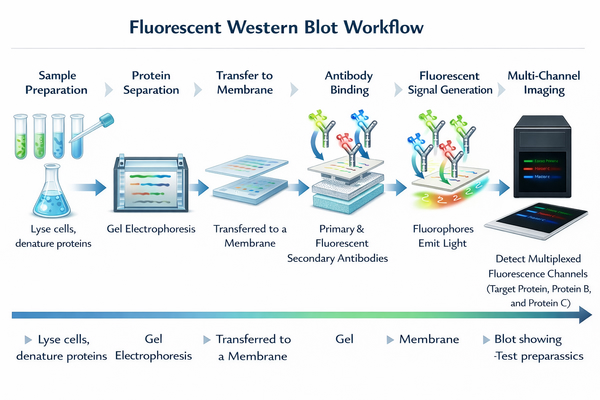 Fig. 2. Fluorescent western blot workflow (BOC Sciences Authorized).
Antibody–Antigen Binding and Fluorescent Signal Generation
Specific binding between antibody and target protein remains the basis of signal generation. Unlike chemiluminescence, the fluorescent signal is directly produced by dye molecules conjugated to antibodies under specific excitation light. Signal intensity correlates well with antibody binding over a wide range, avoiding biases from substrate diffusion, reaction rate differences, or exposure variations. For quantitative analysis, multiple scans of the same membrane can be performed without adjusting imaging parameters, allowing validation of signal stability. The non-destructive nature of fluorescence enables subsequent analyses or archiving, supporting data traceability.
Direct vs. Indirect Fluorescent Antibody Labeling Strategies
Fluorescent Western Blot can use direct or indirect labeling strategies, each with distinct advantages in sensitivity, background control, and experimental complexity. Direct labeling attaches the dye to the primary antibody, generating a signal immediately upon binding. This reduces steps and nonspecific binding, ideal for multiplexing and high-precision quantification. However, signal intensity is limited by primary antibody binding capacity. Indirect labeling uses an unlabeled primary antibody and a fluorescent secondary antibody, amplifying the signal as multiple secondary antibodies bind each primary. This is advantageous for low-abundance proteins but requires careful consideration of cross-reactivity and background. Researchers often choose or combine strategies based on protein abundance, channel numbers, and throughput needs for optimal results.
Integration with Common Fluorescence Imaging Systems
Successful fluorescent Western Blot requires proper integration with imaging hardware. Modern platforms support multiple excitation sources and channels, efficiently accommodating fluorescent-labeled antibodies with different wavelength ranges. During workflow design, factors such as dye excitation/emission, filter configuration, and imaging sensitivity must be considered to ensure signal separation and consistency across channels. Proper setup reduces crosstalk and improves multiplexing accuracy. Fluorescence imaging also supports digital signal acquisition and quantitative analysis software, upgrading Western Blot from image-based interpretation to data-driven analysis.
Fluorescent-Labeled Antibody Solutions for Different Research Scenarios
Fluorescent-labeled antibodies are more than just an upgrade to detection tools—they represent a versatile analytical solution that can be tailored to diverse research goals. By carefully selecting antibody types, labeling strategies, and detection channel combinations, fluorescent Western Blot can precisely match complex research scenarios, from basic expression analysis to system-level pathway studies, significantly enhancing data depth and reliability.
Quantitative Protein Expression Analysis
In quantitative protein expression studies, researchers often need to compare subtle differences in protein levels across samples or treatment conditions. Traditional detection methods, limited by dynamic range and signal saturation, can mask true expression differences. Fluorescent-labeled antibodies provide stable and linearly quantifiable signals, offering higher accuracy in relative quantification. In these applications, fluorescent Western Blot can simultaneously detect target and reference proteins under the same imaging conditions, minimizing systematic errors during normalization. This is especially critical for experiments requiring precise comparisons, such as gene knockdown, overexpression validation, or response analysis after stimulation.
Comparative Studies Across Treatment Conditions
In drug screening, treatment optimization, or time-course studies, the focus is often on expression trends rather than absolute signal intensity at a single time point. Fluorescent-labeled antibodies enable parallel detection of multiple samples and targets on the same membrane, ensuring high comparability across conditions. Multi-channel fluorescence allows researchers to obtain expression changes for multiple proteins in a single experiment, reducing operational errors and batch variability. This parallel detection mode not only improves experimental efficiency but also makes cross-condition comparisons more straightforward and reliable.
Pathway and Signaling Network Analysis
Pathway and signaling network studies typically involve multiple functionally related proteins with expression and activation that are time- and condition-dependent. Single-target detection fails to capture dynamic pathway changes comprehensively. Fluorescent-labeled antibodies provide multiplexing capabilities, allowing simultaneous analysis of multiple key proteins on the same membrane. By carefully designing dye channels and antibody combinations, researchers can monitor upstream, midstream, and downstream proteins, enabling a more complete understanding of signal transduction and regulatory mechanisms. This multi-dimensional data acquisition is valuable for constructing robust pathway models and validating hypotheses.
High-Throughput and Multiplex Western Blot Experiments
In high-throughput studies or large-scale sample screening, efficiency and data consistency are critical. Fluorescent Western Blot simplifies workflows by reducing stripping and repeated incubation steps, making it ideal for high-throughput applications. Multiplex detection allows more information to be obtained in a single experiment, reducing sample consumption and costs. Digital fluorescence acquisition supports automated analysis and data integration, enabling a standardized, scalable Western Blot system. For research teams handling large sample volumes within limited time, fluorescent-labeled antibodies enhance both detection capability and experimental flexibility.
Key Technical Parameters for Optimized Fluorescent Western Blot Performance
Although fluorescent Western Blot offers significant advantages in signal stability and multiplexing, its performance depends heavily on the careful design and coordinated optimization of key technical parameters. Systematic control of these factors is essential to ensure experimental sensitivity, quantitative accuracy, and reproducibility.
Fluorophore Brightness, Stability, and Spectral Separation
The physical properties of fluorophores directly affect detection quality. High-brightness dyes produce sufficient signals under low protein load, reducing background interference and expanding the detectable range. Good photostability prevents signal loss during repeated scans or long imaging sessions, ensuring data consistency. In multiplex experiments, spectral separation is critical. Fluorophores must have sufficient excitation/emission differences to avoid channel crosstalk. Well-designed dye combinations improve multi-channel detection accuracy and provide a reliable foundation for subsequent quantitative analysis.
Antibody Specificity and Cross-Reactivity Control
Antibody specificity remains a core determinant of Western Blot reliability. In fluorescent systems, non-specific binding can be more readily detected by high-sensitivity imaging, amplifying background noise. Careful selection of antibody sources, optimization of conjugation ratios, and appropriate blocking and washing conditions can effectively reduce cross-reactivity. Species- and isotype-specific control is particularly important in multiplex assays, preventing interference between antibodies and ensuring clear, multi-channel signals.
Compatibility with Membranes, Buffers, and Imaging Hardware
Fluorescent Western Blot is a system that involves membrane materials, buffer composition, and imaging devices. Different membranes vary in background fluorescence and signal retention, and buffer components can affect fluorophore stability and antibody binding. During experimental design, it is essential to consider labeling strategy, membrane selection, and imaging system parameters. Systematic optimization of these factors enhances detection sensitivity and minimizes biases from hardware or material incompatibility.
Primary and Secondary Fluorescent-Labeled Antibody Options
Choosing appropriate primary or secondary fluorescent-labeled antibodies is key to detection sensitivity, multiplexing capability, and experimental complexity. Different types of fluorescent antibodies offer distinct advantages in signal strength, background control, and flexibility, and should be selected according to specific research goals.
Fluorescent-Labeled Primary Antibodies for Direct Detection
Fluorescent-labeled primary antibodies have the dye directly conjugated to the antibody that recognizes the target protein, producing a signal upon binding. This direct detection simplifies workflows by reducing incubation and washing steps, minimizing potential non-specific binding and background. In multiplex Western Blot, labeled primary antibodies are advantageous: different targets can be simultaneously incubated with antibodies labeled with distinct fluorophores, avoiding secondary antibody cross-reactivity. This is ideal for high-precision quantification and multi-channel detection. However, signal intensity depends on primary antibody affinity and binding quantity, requiring careful optimization for low-abundance proteins.
Fluorescent-Labeled Secondary Antibodies for Signal Amplification
Fluorescent-labeled secondary antibodies are widely used due to their signal amplification capability. Multiple secondary antibodies bind a single primary antibody, enhancing fluorescence and improving sensitivity for low-expression proteins. This indirect approach is suitable for routine Western Blot and exploratory studies and is cost-effective. In multiplex experiments, careful design of species and isotype specificity is necessary to prevent signal overlap and cross-reactivity between targets. Proper secondary antibody planning is essential for high-quality multi-channel fluorescence detection.
Species-Specific and Isotype-Specific Antibody Design
In complex or multiplex systems, species- and isotype-specific design is crucial for background control and signal clarity. Selecting fluorescent antibodies targeting specific host species and immunoglobulin isotypes reduces off-target binding and enhances signal accuracy. In co-incubation experiments, pairing primary antibodies from different species with highly specific fluorescent secondaries ensures clear, non-interfering multi-channel detection. This approach improves experimental success rates and provides greater flexibility for complex protein analyses.
Custom Fluorescent Antibody Labeling Services at BOC Sciences
BOC Sciences offers professional custom fluorescent antibody labeling services, providing researchers with efficient, precise, and reliable antibody detection and imaging solutions. Our team combines advanced conjugation chemistry, extensive experimental experience, and strict quality control to customize labeling strategies according to research goals, ensuring optimal antibody function and fluorescence performance. Whether for basic research or complex applications, BOC Sciences provides comprehensive technical support and flexible custom solutions.
Diverse Fluorophore Selection
- FITC, Rhodamine, Alexa Fluor series, and other dyes for basic imaging to high-end multiplex experiments.
- Optimized dye selection according to antibody type, application, and experimental conditions for maximum signal and contrast.
- Support for multiplex labeling to allow a single antibody to work across multiple detection channels.
- Fluorescence performance evaluation and reporting to guide optimal dye choice and improve experimental success.
Precise Antibody Conjugation Techniques
- Advanced covalent conjugation ensures stable dye attachment while preserving antibody biological activity.
- Optimal labeling site design for monoclonal, polyclonal, and recombinant antibodies to avoid interfering with antigen binding.
- Post-conjugation functional and fluorescence verification ensures reliable use in experiments.
- Capable of handling special or sensitive antibodies for high conjugation success rates.
Customized Optimization Services
- Flexible adjustment of dye-to-antibody ratios to optimize signal and signal-to-noise ratio.
- Stability optimization under various storage and usage conditions to maintain performance throughout experiments.
- Small-scale trial labeling for pre-experiment validation, reducing risk and cost.
- Personalized strategies for special applications, such as low-abundance proteins or high-background tissues, to ensure accurate results.
Fluorescent Western Blot Support
- Fluorescent-labeled antibodies specifically designed for Western Blot to enhance sensitivity and visualization.
- Support for multi-channel imaging for simultaneous multi-protein detection, improving efficiency and data consistency.
- Stable and reproducible fluorescence signals, unaffected by long-term storage or multiple scans.
- Post-labeling technical support, including signal optimization advice and imaging parameter guidance, ensuring smooth experimental execution.
Do You Need A Consultation?
BOC Sciences integrates cutting-edge fluorescence technologies to accelerate your research, driving next-generation solutions for drug discovery and diagnostics.
Transform Your Studies with Cutting-Edge Fluorescent Products
| Catalog | Name | CAS | Inquiry |
|---|---|---|---|
| F04-0033 | 5-Aminofluorescein | 3326-34-9 | Bulk Inquiry |
| A16-0170 | Rhodamine-123 | 62669-70-9 | Bulk Inquiry |
| A19-0040 | Hoechst 33342 | 23491-52-3 | Bulk Inquiry |
| F06-0011 | Coumarin 153 | 53518-18-6 | Bulk Inquiry |
| F02-0026 | Cy5-NHS ester | 146368-14-1 | Bulk Inquiry |
| F01-0221 | BODIPY Green 8-P2M | 929679-22-1 | Bulk Inquiry |
| A16-0201 | DAPI dihydrochloride | 28718-90-3 | Bulk Inquiry |
| A17-0186 | Perylene Orange | 82953-57-9 | Bulk Inquiry |
| A19-0103 | SYBR Green I | 178918-96-2 | Bulk Inquiry |
| F01-0251 | BODIPY 576/589 | 150173-78-7 | Bulk Inquiry |
High-Performance Fluorescent Tools for Your Research
- Cyanine3 Standard green-orange fluorescent biomolecular labeling.
- Cyanine3.5 Orange-red fluorescence for multiplex imaging.
- sulfo-Cyanine5.5 Water-soluble far-red imaging probe.
- Cyanine7.5 Extended NIR imaging for in vivo studies.
- sulfo-Cyanine Water-soluble cyanine dyes for labeling.
- sulfo-Cyanine3 Hydrophilic green-orange fluorescent conjugation.
- Alexa Fluor Bright, photostable dyes for fluorescence imaging.
- Cyanine Versatile fluorophores for bioimaging applications.
Explore More Topics
Online Inquiry
