

Immunofluorescence Staining
In various life science research fields such as molecular biology, pathology, and drug development, immunofluorescence staining (IF) has become an essential tool for observing the localization and expression levels of target proteins in cells and tissues. As a professional provider of fluorescent dyes and probes, BOC Sciences leverages its deep expertise in fluorescence chemistry, antibody conjugation, biomarker labeling, and custom services to offer comprehensive immunofluorescence staining solutions. These solutions cover the entire process, from dye screening and probe design to labeling services and quality control, catering to global research and industrial clients. Whether customers are engaged in basic research or translational medicine applications, BOC Sciences offers a wide range of fluorescent dyes and customized labeling services to meet diverse experimental needs, helping clients obtain more accurate and biologically relevant data in high-sensitivity, high-resolution, and multi-channel imaging.
Immunofluorescence Testing
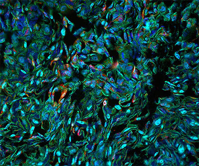
Immunofluorescence staining is a technique that conjugates antibodies with fluorescent dyes to specifically identify and label target antigens, such as organelle proteins, signaling pathway factors, structural proteins, and more. The basic principle involves the use of primary or secondary antibodies to bind to the target protein, emitting a fluorescent signal at specific wavelengths through the fluorescence dye. This allows spatial localization and quantitative analysis under a fluorescence microscope or confocal system. Fluorophores emit longer-wavelength fluorescence signals when excited by specific wavelengths, and these signals can be captured under a microscope. The microscope distinguishes different types of fluorescence against a dark background, forming an image with characteristic peaks. Common fluorophores include FITC and tetramethylrhodamine isothiocyanate (TRITC). Additionally, immunofluorescence staining is often combined with non-antibody-based fluorescent labeling methods, such as using DAPI to stain DNA in the cell nucleus. Immunofluorescence staining has significant advantages in target recognition sensitivity, specificity, spatial resolution, signal enhancement, and data analysis. However, its primary limitation is that it can only be used to observe subcellular structures in fixed (i.e., dead) cells. Moreover, experimental results can be influenced by various factors, such as the type, quality, and concentration of antibodies used, all of which need to be carefully optimized and validated to achieve ideal signal-to-noise ratios and imaging quality.
Advantages of BOC Sciences in Immunofluorescence Staining Solutions
Wide Range of Dyes with Selectable Performance
BOC Sciences offers a variety of fluorescent dyes spanning the ultraviolet to near-infrared spectrum to meet single-channel, multiplex labeling, and high-sensitivity detection needs.Professional Conjugation and Modification Capabilities
With a mature antibody/protein labeling platform, BOC Sciences can efficiently conjugate dyes with primary antibodies, secondary antibodies, small molecule probes, proteins, etc., according to customer needs.High-Quality Standards and Quality Control Services
All dye products undergo rigorous testing for purity, spectral characteristics, fluorescence intensity, and other indicators to ensure experimental stability and reproducibility.Flexible Customization Services to Meet Research Needs
Custom services are available, ranging from dye design, functional group modification to large-scale dye supply, supporting the development of novel fluorescent probes and labeling schemes.
Fluorescent Dye Solutions for Immunofluorescence Staining by BOC Sciences
BOC Sciences has years of expertise in the development of fluorescent dyes and probes, dedicated to providing a complete set of high-performance fluorescent dye solutions for immunofluorescence staining for global research and industrial clients. Combining in-house synthesis capabilities with advanced conjugation technologies, we offer a rich product matrix including classical dyes, high-performance dyes, nuclear dyes, and control dyes to meet various application needs from basic research to high-throughput screening. These fluorescent dyes offer high photostability, good water solubility, and excellent conjugation compatibility with antibodies and biomolecules. They can be widely used in cell labeling, tissue imaging, multiplex immunodetection, colocalization experiments, and other high-resolution imaging technologies. BOC Sciences' fluorescent dye products not only cover the ultraviolet to near-infrared spectrum but also support sensitivity optimization and multi-color imaging design, providing comprehensive support for immunofluorescence staining experiments.
Conventional Fluorophores
Advanced Performance Dyes
- Alexa Fluor series
- CF® Dyes series
- DyLight™ series
- Atto Dyes
Nuclear & Control Dyes
- 4',6-diamidino-2-phenylindole (DAPI)
- Hoechst 33342/Hoechst 33258
- Propidium Iodide (PI)
- Sytox® Green/Sytox® Blue
- MitoTracker® series
Antibody Labeling and Probe Customization Services for Immunofluorescence Staining
In precise, high-throughput immunofluorescence staining experiments, standardized dyes are often insufficient to meet the complex requirements of all research scenarios. To address this, BOC Sciences has expanded its capabilities from basic dye supply to advanced customized services, establishing a one-stop platform for antibody labeling, small molecule probe conjugation, functional group modification, and industrial-scale dye packaging. By combining advanced conjugation chemistry and rigorous quality control processes, we provide high-purity, high-conjugation efficiency, and low-background interference customized products, widely applicable in basic research, pathological analysis, drug screening, diagnostic development, and other fields. Whether constructing specific fluorescent primary/secondary antibodies, designing small molecule probes for live cell imaging, or developing large-scale dye solutions for automation systems, BOC Sciences offers flexible, efficient, and scalable solutions to help customers achieve more complex multi-color imaging and molecular recognition tasks.
Custom Fluorescent Labeling of Primary/Secondary Antibodies
We select the most suitable fluorescent dyes for covalent conjugation based on the customer-provided antibody specifications, ensuring both labeling efficiency and biological activity. Customization is supported for common antibody species (rabbit, mouse, human, etc.) and various dye combinations.
Fluorescent Conjugate Probe Design
For special target detection and live cell imaging needs, BOC Sciences offers customized fluorescent conjugation services for small molecule probes, peptide probes, proteins, or enzyme probes.
Functional Group Modification Services
We provide dye modifications with multifunctional groups such as biotin, azide (N₃), alkyne, maleimide, and carboxyl, suitable for applications like click chemistry, crosslinking reactions, and affinity purification.
Bulk Packaging and Industrial-Scale Supply
For high-throughput platforms, diagnostic companies, and industrial applications, we offer dye products in gram, 10g, and 100g quantities, accompanied by COA and inter-batch consistency guarantees, facilitating the transition from laboratory to clinical/industrial applications.
Strict Quality Control Testing System for Fluorescent Dyes
BOC Sciences considers quality control to be the core part of our product development and delivery system, building a dual-standard testing and release system that meets both research-grade and industrial-grade requirements. We are equipped with advanced analytical platforms and an experienced quality analysis team, implementing multi-dimensional testing schemes covering dye purity, labeling efficiency, optical performance, structural confirmation, stability validation, and more, ensuring that each batch performs consistently and reliably in high-complexity experimental environments. Through our continually optimized quality control processes and global customer feedback mechanisms, we enhance product consistency and reproducibility, providing reliable fluorescent dye solutions for life sciences research and in vitro diagnostics.
- Purity Testing: We use HPLC, UPLC, and other methods to determine the purity of the dye's main peak, ensuring it is ≥95% or higher, and controlling by-products and unreacted intermediates.
- Excitation/Emission Spectra Testing: We measure excitation and emission wavelengths, fluorescence intensity, and quantum yield to assess the dye's luminous performance under different environments.
- Degree of Labeling (DOL) Testing: We calculate the labeling degree of conjugated antibodies or probes to ensure the balance between signal strength and molecular functionality.
- Molecular Weight and Structure Validation: Using mass spectrometry (MS) and nuclear magnetic resonance (NMR), we confirm the dye's structure and integrity to avoid synthetic deviations.
- Stability Testing: We assess dye storage stability and performance retention under various conditions of temperature, light, and pH to ensure consistent performance during transport and long-term storage.
- Solubility and Buffer Compatibility Testing: We test the dye’s solubility in different pH and buffer systems to ensure its operability in cell staining and tissue immunoassays.
Comprehensive Detection Instruments
- High-Performance Liquid Chromatograph (HPLC)
- Ultra-Performance Liquid Chromatograph (UPLC)
- UV-Visible Spectrophotometer (UV-Vis)
- Fluorescence Spectrophotometer
- Mass Spectrometer (MS, LC-MS)
- Nuclear Magnetic Resonance Spectrometer (NMR)
- Dynamic Light Scattering (DLS)
- Stability Chamber
- pH/Buffer Compatibility Testing Devices
- Automated Microscopic Imaging Platform
Immunofluorescence Staining Research Supported by Our Fluorescent Dye Solutions
BOC Sciences' fluorescent dye solutions are widely applied in various immunofluorescence staining studies, covering fields such as cell biology, pathology, cancer research, viral and infectious disease research, and drug screening and mechanism studies. Our products undergo strict quality control to ensure excellent performance under various experimental conditions. Whether for basic research, preclinical studies, or industrial applications, BOC Sciences provides efficient and reliable fluorescent dye support to help customers explore biological processes, disease mechanisms, and drug action mechanisms in-depth.
Cell Biology Research
Immunofluorescence staining is widely used in cell biology research to observe cell structure, locate organelles, and analyze intracellular signaling. BOC Sciences' highly sensitive fluorescent dyes help researchers accurately detect and track key molecules inside cells, studying processes like cell proliferation, differentiation, and migration.
Pathology Research
In pathology research, immunofluorescence staining is used for molecular labeling of pathological tissue sections to help identify diseased tissues and early cancer diagnosis. Using fluorescent dyes of different wavelengths, researchers can perform multiplex labeling on the same section, improving detection sensitivity and accuracy.
Cancer Research
In cancer research, immunofluorescence staining is mainly used for detecting tumor markers and analyzing the tumor microenvironment. The dyes provided by BOC Sciences can label characteristic molecules of tumor cells, helping researchers better understand the cancer development process and evaluate potential targeted therapies.
Viral and Infection Research
BOC Sciences' fluorescent dyes assist researchers in tracking pathogen invasion processes in viral and infection studies. Using fluorescent immunostaining, researchers can observe the interaction between viruses and host cells in real-time, uncovering infection mechanisms and transmission pathways.
Drug Screening and Mechanism Research
Immunofluorescence staining is used in drug screening and mechanism studies for high-throughput screening, toxicity evaluation, and drug action mechanism analysis. By using fluorescent dyes to label target molecules or drugs, BOC Sciences’ dye solutions provide critical information for drug development, helping identify potential therapeutic agents and effective components.
Frequently Asked Questions
-
How does immunofluorescence staining work?
Immunofluorescence staining is a technique used to detect specific proteins or antigens in cells or tissues. It involves using antibodies that are conjugated to a fluorescent dye, which binds specifically to the target antigen. The process starts by fixing the sample to preserve its structure. Then, primary antibodies are applied to the sample, followed by secondary antibodies that are tagged with a fluorescent dye. When the sample is illuminated with a specific wavelength of light, the dye emits fluorescence, allowing visualization of the target antigen under a microscope. Different fluorescent dyes can be used for multicolor staining to detect multiple targets simultaneously.
-
How to do immunofluorescence staining?
To perform immunofluorescence staining, first, prepare the sample by fixing cells or tissue sections using formaldehyde or another fixative. After fixation, permeabilize the cells using a detergent like Triton X-100 to allow antibody access. Block non-specific binding sites with a blocking solution such as bovine serum albumin (BSA). Then, incubate the sample with the primary antibody specific to the target protein, followed by washing to remove unbound antibodies. Next, apply a secondary antibody conjugated to a fluorescent dye, incubate, and wash again. Finally, mount the sample on a microscope slide with a mounting medium, and visualize the fluorescence under a confocal or fluorescence microscope.
-
How to quantify immunofluorescence staining?
Quantifying immunofluorescence staining typically involves measuring the intensity of fluorescence signals. This can be done using software tools that analyze images captured by a fluorescence microscope. The intensity of fluorescence is proportional to the amount of the target antigen present. The process usually involves selecting regions of interest (ROIs), calculating the fluorescence intensity within those areas, and normalizing the signal to factors like cell size or background intensity. Advanced methods, such as flow cytometry or image-based analysis, can also be used to quantify staining in a more automated and high-throughput manner, providing more accurate and reproducible results.
-
How to reduce background staining in immunofluorescence?
Reducing background staining in immunofluorescence involves several strategies. First, ensure proper blocking to prevent non-specific antibody binding, using solutions like BSA or serum from the same species as the secondary antibody. Second, optimize antibody concentrations, as too much antibody can lead to excess binding. Additionally, using high-quality, well-characterized primary and secondary antibodies can minimize non-specific interactions. Washing the sample thoroughly after each antibody incubation step is crucial to remove unbound antibodies. Finally, using high-quality mounting media and ensuring proper sample fixation and permeabilization conditions can help reduce background fluorescence and improve signal clarity.
High-Performance Fluorescent Tools for Your Research
- Cyanine3 Standard green-orange fluorescent biomolecular labeling.
- Fluorescent Dyes General-purpose labeling for bioanalytical detection.
- Cyanine5.5 Far-red fluorescence for deep tissue imaging.
- TAMRA Dyes Red fluorescent labeling for biomolecule tracking.
- Cyanine7.5 Extended NIR imaging for in vivo studies.
- JOE Dyes Green fluorescent probes for qPCR applications.
- Alexa Fluor Bright, photostable dyes for fluorescence imaging.
- BODIPY Photostable dyes for lipid and cell imaging.
Fluorescent Services
-

Fluorescent Beads
Bright, uniform microspheres for calibration and tracing.
-

Fluorescent Nanoparticles
Stable, bright nanoscale probes for imaging and sensing.
-

Fluorescent Proteins
Genetically encoded fluorescent markers for live-cell imaging.
Explore More Topics
Online Inquiry
