

Rhodamine
-

-

-

-
-

-

-
 Rho110 NHS Ester
CAT:
-
 Rho101 NHS Ester
CAT:
-
 9-[2-Carboxy-5(or 6)-isothiocyanatophenyl]-3,6-bis(diethylamino)xanthylium, chloride
CAS No.:
Purity:
CAT:
-

-

-

-
 ROX amine, 6-isomer
CAT:
-

-

-
 ATT 594 azide
CAT:
-
 ATT 565 azide
CAT:
-
-
-
Background
Rhodamine is a class of high-brightness fluorescent dyes with excellent photostability, widely used in biological labeling, cell imaging, microbial detection, and materials science research. Compared with other commonly used fluorescent dyes, Rhodamine dyes possess advantages such as strong photostability, pH insensitivity, high extinction coefficients, longer absorption and emission wavelengths (generally >500 nm), and high fluorescence quantum yields. Therefore, Rhodamine enables efficient labeling of proteins, nucleic acids, lipids, and antibodies, while supporting multichannel fluorescence imaging in both live and fixed cells. Its unique optical properties and broad applications make Rhodamine an indispensable fluorescent tool in scientific and industrial fields, providing reliable support for experimental design and data analysis. BOC Sciences is dedicated to providing customers with high-quality Rhodamine dyes and their derivatives. We not only supply a comprehensive range of Rhodamine compounds covering products suitable for different chemical functional groups (such as carboxyl, amino, azide, and maleimide), but also design customized structures tailored to specific experimental needs. Our products feature high purity and high stability, ensuring reliable performance under complex experimental conditions.
What is Rhodamine Dye and Its Key Features?
Rhodamine dyes are an important class of triphenylmethyl-based fluorescent dyes, widely applied in life sciences, chemical analysis, and materials science research. Their core structure consists of an aromatic ring system substituted with amino and carboxyl groups, which confers extremely high absorption and emission efficiency in the visible light range. The chemical stability and conjugated system of Rhodamine not only provide bright and long-lasting fluorescence signals but also enable convenient functionalization of derivatives. With tunable optical properties, Rhodamine has become an indispensable tool in experiments such as protein labeling, nucleic acid detection, and cell imaging.
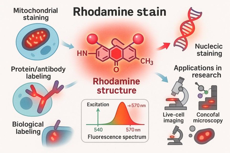 Fig. 1. Rhodamine stain (BOC Sciences Authorized).
Rhodamine Structure and Chemical Properties
Rhodamine dyes are generally based on a triphenylmethyl backbone, containing different substituents to adjust their optical and chemical characteristics. Typical structures include Rhodamine 123, Rhodamine B, and their derivatives. Rhodamine exhibits a strong conjugated system, giving it a high molar absorption coefficient and significant fluorescence signal in the visible range. In terms of chemical properties, Rhodamine can undergo modifications such as acylation, etherification, and PEGylation, thereby improving its water solubility, membrane permeability, and biocompatibility.
Rhodamine Solubility in Different Solvents
Rhodamine dyes show distinct solubility differences in various solvents. They dissolve well in polar solvents such as water, methanol, and ethanol, but have limited solubility in non-polar solvents. To meet the needs of biological experiments and materials science studies, researchers often use PEGylated or ionized derivatives to enhance water solubility and stability. This not only facilitates the preparation of uniform fluorescent Rhodamine solutions but also reduces precipitation or uneven distribution during cell or tissue staining experiments, ensuring reproducibility and accuracy of results.
Understanding Rhodamine Spectrum: Excitation and Emission
When selecting an appropriate Rhodamine dye, it is crucial to understand its excitation and emission characteristics, quantum yield, and fluorescence color. Different derivatives exhibit significant differences in excitation and emission wavelengths, while quantum yield directly affects fluorescence brightness and signal stability. The emission color determines the dye's distinguishability in multiplex labeling experiments. Therefore, mastering the spectral properties of Rhodamine is key to experimental design and optimization of results.
| Rhodamine Dye | Excitation Wavelength (nm) | Emission Wavelength (nm) | Quantum Yield | Fluorescence Color | Key Application Features |
| Rhodamine B | 540 | 625 | 0.65 | Red | Cytoplasmic staining, multicolor labeling, microscopy imaging |
| Rhodamine 6G | 530 | 550 | 0.95 | Bright Green | High-sensitivity detection, strong photostability, widely used in industry |
| Rhodamine 123 | 507 | 529 | 0.90 | Green | Mitochondrial staining, live-cell imaging, membrane potential studies |
| Rhodamine 110 | 496 | 520 | 0.88 | Green | Protein and nucleic acid labeling, good water solubility |
| Tetramethylrhodamine (TMR) | 555 | 580 | 0.66 | Orange-Red | Antibody conjugation, cytoplasm/membrane labeling, good photostability |
| Carboxytetramethylrhodamine (TAMRA) | 559 | 583 | 0.63 | Orange-Red | Carboxyl-functionalized, peptide/protein labeling, water-soluble |
| Sulforhodamine 101 | 586 | 605 | 0.90 | Red | Live-cell staining, neural imaging, excellent water solubility |
Notes:
- Excitation and emission wavelengths can be used to select appropriate light sources and filters to ensure clear fluorescence signals.
- The higher the quantum yield, the brighter the fluorescence signal, making it suitable for highly sensitive detection.
- Fluorescence color determines the distinguishability in multiplex labeling experiments.
- Application features help researchers quickly choose the right Rhodamine dye for their experimental purposes.
Optimization of Photostability of Rhodamine Probes
The photostability of Rhodamine dyes is critical to experimental reliability, especially in live-cell imaging or long-term fluorescence detection. Photobleaching may cause signal decay and affect data accuracy. By optimizing the chemical structure, such as introducing hydroxyl or ester groups on the aromatic ring, and by adding antioxidants to the experimental system or avoiding prolonged strong light exposure, photostability can be effectively enhanced. In addition, proper selection of filters and laser intensity can also reduce photobleaching effects, thereby extending the lifespan of the dye and ensuring experimental reproducibility.
Rhodamine Synthesis and Custom Molecular Modifications
The chemical synthesis of Rhodamine dyes generally involves multi-step complex reactions, including selective substitution on aromatic rings, cyclization, and various coupling reactions. These reactions not only determine the core structure of the dye but also directly influence its optical properties, solubility, and chemical stability. Furthermore, Rhodamine dyes can undergo various functional modifications, such as PEGylation and conjugation with proteins or polysaccharides, thereby expanding their applications in biological labeling, cell imaging, and materials science. These synthesis and molecular modification strategies provide researchers with abundant tools and flexibility in experimental design.
Rhodamine Synthesis Strategies and Challenges
The synthesis of Rhodamine dyes involves multiple complex chemical reactions, including selective substitution of aromatic rings, cyclization reactions, and various coupling steps. These reactions not only define the core structure of the dye but also directly affect its optical performance, solubility, and chemical stability. At the same time, the chemical synthesis of Rhodamine faces multiple challenges, including selective coupling, purification of reaction products, and maintaining photostability during synthesis. In practice, by optimizing reaction conditions, selecting appropriate solvent systems and catalysts, and controlling temperature and reaction time, the yield and purity of target products can be improved, ensuring the consistency and reliability of Rhodamine dyes in both scientific and industrial applications.
Rhodamine Conjugated Compounds
Due to their high brightness and excellent optical stability, Rhodamine dyes are often covalently conjugated with biomacromolecules or functional molecules to achieve specific experimental and imaging functions. Through chemical conjugation, Rhodamine can be linked to proteins, peptides, polysaccharides, antibodies, and even small molecule ligands, forming derivatives with targeting or tracing abilities. These conjugated molecules not only retain the fluorescence characteristics of Rhodamine but also enhance stability and uniform distribution in aqueous solutions or biological systems. Different conjugation strategies, such as PEGylation, acylation, or biotinylation, can further expand the applications of Rhodamine in cytoskeleton labeling, membrane tracking, nucleic acid detection, and immunofluorescence, offering researchers versatile and flexible tools.
PEGylated Rhodamine
By modification with polyethylene glycol, the water solubility and biocompatibility of the dye are improved, making it suitable for in vivo imaging and live-cell experiments. It can extend blood circulation time and reduce nonspecific binding.
Rhodamine Conjugated Phalloidin
Specifically binds to actin filaments, enabling visualization of cytoskeletal structures and dynamic changes. It is an important tool in cell biology and structural biology research.
Rhodamine Conjugated Dextran
Used to trace intracellular transport pathways and tracking experiments. By conjugating with polysaccharides, the dye can remain stable inside cells and avoid rapid efflux, thereby improving experimental reproducibility.
Rhodamine Conjugated Streptavidin
Capable of selectively binding to biotinylated molecules, enabling fluorescent labeling and detection of proteins, nucleic acids, or other biomacromolecules. It is widely applied in immunofluorescence, flow cytometry, and molecular diagnostic experiments.
Looking for Expert Guidance?
BOC Sciences harnesses cutting-edge Rhodamine dye technologies to accelerate your research and drive innovation in imaging, sensing, and diagnostics.
Comparing Different Rhodamine Derivatives for Optimal Use
Rhodamine dye derivatives are diverse, and through structural and functional group modifications, they can meet various experimental conditions and research requirements. Different derivatives exhibit significant differences in reactivity, water solubility, optical performance, and conjugation methods with biomacromolecules. Understanding these differences is essential for selecting the appropriate Rhodamine dye, which can improve labeling efficiency, enhance the stability of fluorescence signals, and optimize applications in cell imaging, protein or nucleic acid labeling, and materials science research.
| Rhodamine Derivative | Functional Group | Key Features | Typical Applications |
| Rhodamine Acrylate | Acrylate | Can form covalent bonds with polymers via radical polymerization; stable optical properties. | Fluorescent polymer labeling, polymer material staining. |
| Rhodamine Alkyne | Alkyne | Suitable for Click chemistry conjugation; high reaction specificity. | Protein, nucleic acid, and small molecule labeling. |
| Rhodamine Amine | Amine | Contains active amino group; good water solubility; reacts with activated carboxyl groups. | Protein labeling, cell staining, various conjugation experiments. |
| Rhodamine NHS Ester | N-hydroxysuccinimide (NHS) | Highly selective; reacts efficiently with amino groups on proteins or peptides. | Protein labeling, antibody conjugation, immunofluorescence. |
| Rhodamine Maleimide | Maleimide | Forms covalent bonds with cysteine thiol groups; high specificity. | Protein thiol labeling, live-cell imaging. |
| Rhodamine Carboxylic Acid | Carboxyl | Good water solubility; can be activated to NHS ester or conjugated to polysaccharides/proteins. | Multi-molecule conjugation, fluorescence tracing. |
| Rhodamine Sulfonate | Sulfonate | Extremely water-soluble; negatively charged; suitable for aqueous systems. | Aqueous imaging, extracellular labeling, cell tracking. |
| Rhodamine Biotin Conjugate | Biotin | Can bind streptavidin with high selectivity. | Immunodetection, multi-molecule complex tracing. |
| Rhodamine Dextran Conjugate | Dextran | Large molecule carrier; prolongs intracellular retention. | Intracellular transport studies, vascular or neural tracing. |
| Rhodamine PEGylated | Polyethylene glycol (PEG) | Improves water solubility and biocompatibility. | In vivo imaging, drug carrier tracking. |
Expert Rhodamine Labeling Techniques for Biomolecules
Rhodamine dyes, due to their high brightness and excellent photostability, are widely applied in labeling proteins, nucleic acids, lipids, and antibodies. Proper selection of labeling strategies and conditions not only enhances fluorescence signal intensity but also improves detection sensitivity and specificity while significantly reducing background signals and nonspecific binding. By optimizing reaction systems, buffer conditions, and dye structures, researchers can achieve precise and controlled fluorescence labeling of different biomolecules, providing reliable tools for cell imaging, protein interaction analysis, and molecular diagnostics.
Rhodamine Labeling for Proteins
Rhodamine dyes can specifically bind to target proteins through carboxyl activation or amine coupling reactions, achieving efficient fluorescence labeling. For example, rhodamine-labeled fibronectin can be used to study extracellular matrix structures, cell adhesion, and migration mechanisms. By optimizing dye-to-protein molar ratios, reaction time, and pH conditions, labeling efficiency can be significantly improved while preserving protein bioactivity. Such labeled proteins can be applied in confocal microscopy imaging, cell motility tracking, and protein-protein interaction studies.
Rhodamine Labeled Peptide
Rhodamine labeling of short peptides provides powerful tools for studying intracellular signal transduction, protein interactions, and drug delivery pathways. By introducing Rhodamine at the N-terminus or C-terminus of peptides, bioactivity can be maintained while enabling visualization and tracking. In experiments, selecting appropriate buffer systems and dye concentrations, combined with suitable purification methods, reduces nonspecific binding and dye self-aggregation, resulting in high signal-to-noise fluorescence signals suitable for live-cell imaging and in vitro functional analysis.
Rhodamine Antibody Conjugation
Rhodamine-conjugated antibodies are widely used in immunofluorescence staining, flow cytometry, and molecular imaging. By optimizing conjugation ratios, selecting suitable buffer systems, and controlling reaction temperature, high labeling efficiency and low nonspecific binding can be achieved. Different types of Rhodamine derivatives, such as NHS esters or maleimides, can specifically conjugate with antibody amines or thiols, ensuring uniform, bright fluorescence signals and enabling multicolor labeling experiments for complex biological analyses.
Nucleic Acid Labeling Using Rhodamine Dyes
Rhodamine dyes can label DNA and RNA through oligonucleotide end modifications or internal base conjugation. Such labeling is widely applied in fluorescence in situ hybridization (FISH), quantitative real-time PCR (qPCR), and fluorescent probe detection. By selecting suitable derivatives and optimizing labeling conditions, researchers can obtain bright, stable, and high signal-to-noise fluorescence signals, improving nucleic acid detection sensitivity and accuracy while supporting multiplex labeling experiments.
Rhodamine Labeled Phospholipid
Rhodamine-labeled phospholipids are widely used in studies of cell membrane dynamics, vesicle trafficking, and lipid interaction analysis. Through covalent conjugation or lipid insertion strategies, Rhodamine can maintain membrane integrity while providing strong fluorescence signals. These labeled phospholipids can be applied in live-cell imaging, membrane fusion experiments, and lipid microdomain tracking, supporting research on membrane protein distribution, vesicle transport mechanisms, and lipid dynamics.
Advanced Rhodamine Staining Techniques for Cellular and Microbial Research
Rhodamine dyes are extensively used in cellular and microbial research for high-resolution imaging and dynamic tracking experiments. By optimizing dye concentration, staining time, and buffer conditions, high-brightness and low-background staining can be achieved in both live and fixed cells. Different Rhodamine dyes, such as Rhodamine B, Rhodamine 123, and Auramine-Rhodamine combinations, can provide specific fluorescence signals for the cytoplasm, mitochondria, nuclei, and membranes, supporting cell biology, pathogenic microorganism detection, and functional imaging studies.
Rhodamine B Staining Cells for Fluorescence Imaging
Rhodamine B is a widely used cell dye applicable to both live and fixed cells. In live-cell experiments, it can penetrate the cell membrane and label the cytoplasm, facilitating the observation of cell morphology, motility, and intracellular structures. In fixed-cell experiments, optimizing staining concentration and time ensures uniform cytoplasmic signals while preserving cellular integrity. Rhodamine B staining can also be combined with other fluorescent probes for multichannel imaging, enhancing experimental information output.
Rhodamine B Staining Lipid Membranes and Organelles
Rhodamine B and its derivatives can specifically label intracellular lipids and lipid vesicles, making them suitable for lipid metabolism and membrane dynamics studies. By embedding into membranes or conjugating with lipid molecules, Rhodamine dyes allow visualization of lipid distribution, membrane fusion, and vesicle trafficking under microscopy. In drug delivery and lipid metabolism research, this staining technique provides high-resolution, dynamic visualization data, helping to reveal intracellular lipid dynamics.
Rhodamine Staining Bacteria for Detection and Analysis
Rhodamine dyes are employed for bacterial detection and fluorescence imaging, commonly used in Auramine-Rhodamine staining protocols to enhance visualization of pathogens such as Mycobacterium. By selecting appropriate dye concentrations and fixation methods, high-contrast and low-background fluorescence signals can be achieved, facilitating microbial counting, morphology observation, and infection research. In pathogenic detection, antibacterial testing, and environmental microbiology, Rhodamine dyes provide reliable and highly sensitive imaging methods.
Rhodamine Staining Mitochondria in Live and Fixed Cells
Rhodamine 123 and related mitochondrial-specific dyes are widely applied in live-cell mitochondrial imaging. These dyes selectively accumulate in mitochondrial membrane potential regions, allowing real-time observation of mitochondrial morphology, dynamic changes, and potential fluctuations under fluorescence microscopy. This technique is crucial for studies of cellular metabolism, apoptosis, and drug action mechanisms. By optimizing staining concentration, incubation time, and buffer conditions, cell function interference can be minimized while achieving strong mitochondrial fluorescence signals.
Rhodamine Staining Nucleus for Cellular Localization
Rhodamine dyes can also be used for nuclear staining, particularly derivatives that bind DNA. By selecting suitable Rhodamine derivatives and staining conditions, highly specific nuclear signals can be obtained, enabling multichannel imaging alongside other intracellular fluorescent markers. Such staining methods are valuable in studies of the cell cycle, nuclear morphology, apoptosis detection, and microbial nuclear structure visualization, providing powerful fluorescent tools for cell biology and microbiology research.
Comprehensive Overview of Rhodamine Dye Uses
Rhodamine dyes, with their high brightness, excellent photostability, and tunable spectral properties, have broad applications across biology, medicine, and materials science. Their unique fluorescence enables researchers to perform highly sensitive detection, cell imaging, and multiplex labeling experiments. In addition, the diverse functional groups of rhodamine derivatives provide flexible options for labeling proteins, nucleic acids, and lipids, supporting experimental needs ranging from basic research to clinical diagnostics.

Rhodamine for Flow Cytometry Applications
In flow cytometry, rhodamine dyes can be conjugated with antibodies or cell-specific probes to provide high signal-to-noise fluorescence. Their high quantum yield and stable emission spectra allow for more accurate and reliable distinction of cell populations. Rhodamine is widely applied in immunophenotyping, cell cycle studies, and apoptosis detection, enabling rapid and quantitative analyses and serving as an ideal fluorescence tool for high-throughput cellular studies.

Rhodamine in Immunofluorescence Assays
Rhodamine dyes are commonly used for labeling antibodies or other biomolecules in immunofluorescence assays, supporting visualization of cells and tissue structures. With their bright red fluorescence and high photostability, rhodamine is suitable for multicolor colocalization experiments. Optimizing conjugation strategies and staining conditions yields high-specificity, low-background signals, making rhodamine widely applicable in protein distribution analysis, signal pathway studies, and pathological tissue observation.

Rhodamine for Live Cell Imaging
Thanks to their cell membrane permeability and low cytotoxicity, rhodamine dyes are extensively used in live-cell imaging. They can specifically label mitochondria, membrane lipids, and other intracellular structures, enabling dynamic observation and functional analysis. Combined with confocal or super-resolution microscopy, rhodamine provides high-resolution, real-time cellular information, supporting research on metabolism, membrane dynamics, and drug mechanisms of action.

High-Throughput Screening with Rhodamine
Rhodamine dyes are applied in high-throughput screening to rapidly quantify enzyme activities, drug responses, and compound library analyses. Their stable fluorescence signals and high brightness ensure more reliable experimental results. By optimizing dye concentrations and detection conditions, rhodamine significantly enhances screening efficiency and sensitivity, making it a valuable fluorescence tool in drug discovery, enzyme screening, and functional genomics.

Microbial Detection and Environmental Analysis
Rhodamine dyes are used in microbial labeling and detection, including bacteria, fungi, and other pathogens. The auramine–rhodamine staining method is widely applied in Mycobacterium tuberculosis detection. Optimized staining and fixation methods provide high-contrast fluorescence signals, suitable for microbial counting, infection studies, and environmental microbial monitoring, delivering reliable support for clinical diagnostics and ecological research.

Membrane Dynamics and Organelle Tracing
Rhodamine dyes are valuable for studying membrane dynamics, vesicle trafficking, and organelle tracing. Through covalent labeling or membrane embedding strategies, rhodamine can mark structures such as mitochondria, endoplasmic reticulum, and the Golgi apparatus. This supports research on membrane fusion, vesicle transport, and organelle function dynamics, widely applied in cell biology, signal transduction, and drug mechanism studies.
How to Choose the Right Rhodamine Dye for Your Experiments
Selecting the appropriate rhodamine dye is critical to experimental success. Researchers must consider experimental objectives, labeling targets, fluorescence wavelength, photostability, and biocompatibility to ensure optimal labeling and staining performance. Experimental design should also account for instrument factors such as light sources, filter configurations, and sample characteristics, enabling the choice of the most suitable rhodamine derivative for bright, low-background fluorescence signals to support high-sensitivity imaging and quantitative analysis.
Rhodamine vs Other Fluorophores
Compared with commonly used fluorophores such as FITC, Alexa Fluor, and Cy dyes, rhodamine offers higher photostability and quantum yield, making it especially effective in long-term imaging and high-intensity experiments. Additionally, rhodamine provides diverse fluorescence colors, covering emission ranges from green to red. However, compatibility of excitation/emission spectra with experimental equipment must be considered to ensure proper matching of light sources and filters, avoiding spectral overlap and signal interference.
Selecting Based on Fluorescence Spectrum
Choosing a rhodamine dye with excitation wavelengths suited to the light source of the microscope or detector maximizes signal-to-noise ratio and minimizes spectral interference. For example, in multicolor colocalization experiments, derivatives with distinct excitation/emission profiles compared to other fluorophores should be selected to achieve clear multichannel imaging. Taking into account quantum yield, dye stability, and sample properties further optimizes signals while balancing sensitivity and accuracy.
Optimizing for Labeling and Staining Efficiency
Improving rhodamine labeling and staining efficiency requires adjusting dye concentration, buffer system, pH, and reaction time while minimizing nonspecific binding and dye aggregation. Selecting the appropriate derivative type, such as NHS ester, maleimide, or PEGylated rhodamine, enables specific conjugation to different biomolecules. Moreover, optimizing staining conditions and incubation parameters not only enhances fluorescence brightness but also significantly reduces photobleaching, ensuring reliable results in labeling proteins, nucleic acids, lipids, and antibodies.
Frequently Asked Questions
-
Is rhodamine dye toxic?
Rhodamine dyes are generally considered to have low toxicity, but strict safety protocols must still be followed during experimental use. Direct contact with dye powders or high-concentration solutions may cause skin or respiratory irritation, so gloves, goggles, and protective clothing should always be worn. For in vitro experiments, potential effects on cell viability can be reduced by selecting low-toxicity derivatives or controlling dye concentration. Proper waste disposal and dye storage are also essential for laboratory safety, helping to minimize environmental contamination and operational risks.
-
What is Rhodamine 6G?
Rhodamine 6G is a bright green-to-yellow fluorescent dye with high quantum yield and excellent photostability. It is commonly used in lasers, fluorescent labeling, optical sensors, and cell imaging. With an excitation wavelength of about 530 nm and an emission wavelength of 550–560 nm, it is a high-performance fluorescent dye frequently employed in both research and industrial applications.
-
What is Rhodamine Red?
Rhodamine Red refers to a class of red fluorescent dye derivatives with emission spectra ranging from 570 to 610 nm. It is commonly used in protein and nucleic acid labeling as well as immunofluorescence assays. With strong photostability and high brightness, Rhodamine Red is well-suited for multicolor imaging and colocalization studies, providing a reliable fluorescence tool for cell biology and molecular imaging.
-
What is Auramine Rhodamine?
Auramine–Rhodamine is a combined staining method used in microbial detection, particularly for fluorescent staining of Mycobacterium tuberculosis. Auramine provides green fluorescence, while rhodamine enhances the red signal. Together, they achieve high-contrast, sensitive detection, widely applied in pathogenic microorganism identification and environmental microbiological analysis.
-
What is the color of rhodamine?
Rhodamine dyes typically exhibit bright fluorescence ranging from orange-red to pink or greenish-yellow, depending on the specific derivative. Their vivid color makes them easily detectable in biological imaging and labeling experiments.
-
What is rhodamine color used for?
The rhodamine color is used to distinguish different targets in multicolor fluorescence experiments, select compatible filters and light sources, and ensure high-contrast, clear imaging in applications such as protein, nucleic acid, or cell labeling.
-
What is the principle of rhodamine assay?
A rhodamine assay relies on the dye's fluorescence intensity to quantify biomolecules, monitor enzyme activity, or track cellular processes. Excitation of the rhodamine molecule produces a strong emission signal, which can be measured to determine concentration or activity with high sensitivity.
Fluorescent Services
-

Fluorescent Beads
Bright, uniform microspheres for calibration and tracing.
-

Fluorescent Nanoparticles
Stable, bright nanoscale probes for imaging and sensing.
-

Fluorescent Proteins
Genetically encoded fluorescent markers for live-cell imaging.
Explore More Topics
Resources

- Hoechst Dyes: Definition, Structure, Mechanism and Applications
- Mastering the Spectrum: A Comprehensive Guide to Cy3 and Cy5 Dyes
- Fluorescent Probes: Definition, Structure, Types and Application
- Fluorescent Dyes: Definition, Mechanism, Types and Application
- Coumarin Dyes: Definition, Structure, Benefits, Synthesis and Uses
- Unlocking the Power of Fluorescence Imaging: A Comprehensive Guide
- Cell Imaging: Definitions, Systems, Protocols, Dyes, and Applications
- Lipid Staining: Definition, Principles, Methods, Dyes, and Uses
- Flow Cytometry: Definition, Principles, Protocols, Dyes, and Uses
- Nucleic Acid Staining: Definition, Principles, Dyes, Procedures, and Uses
Online Inquiry
