

Rhodamine 123 Fluorescent Staining Protocols
Rhodamine 123 (Rh123) is a widely used fluorescent dye in cell biology research that selectively accumulates in the mitochondria of living cells. This property makes it an essential tool for studying mitochondrial function and assessing cell viability. Rh123 has unique chemical characteristics as a lipophilic cationic dye, allowing it to easily penetrate cell membranes and accumulate within mitochondria driven by the mitochondrial membrane potential. Optically, Rh123 is excited at a wavelength of 488 nm and emits a strong green fluorescence at 530 nm. Due to its high quantum yield, Rh123 produces high-contrast fluorescent signals, making it especially valuable for cellular biology experiments.
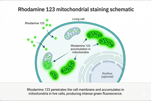 Fig. 1. Rhodamine 123 mitochondrial staining schematic within a cell (BOC Sciences Authorized).
Experimental Principles
Staining Mechanism
The widespread use of Rh123 for studying mitochondrial function is largely due to its specific staining mechanism. Rh123 passively diffuses through the cell membrane into the cytoplasm and is selectively accumulated by mitochondria because of the mitochondrial membrane potential. This accumulation depends on mitochondrial integrity and active membrane potential. Specifically, mitochondria with higher membrane potential accumulate more Rh123, resulting in stronger fluorescence, whereas lower membrane potential leads to reduced accumulation and weaker fluorescence signals.
Experimental Objectives
Fluorescent staining with Rh123 allows researchers to:
- Assess cell viability: Strong Rh123 fluorescence in living cells indicates intact mitochondrial function, providing a measure of cellular viability.
- Evaluate mitochondrial function: Fluorescence intensity within mitochondria reflects the mitochondrial membrane potential and functional state.
- Drug research: Changes in Rh123 fluorescence before and after treatment can assess drug effects on mitochondrial function.
- Metabolic and disease studies: Rh123 can be combined with other markers (e.g., ROS probes or apoptosis indicators) for multiparameter analysis.
Experimental Materials and Equipment
Core Reagents
- Rhodamine 123 solution: Typically prepared as a stock solution and diluted according to experimental requirements. High-purity Rh123 ensures consistent staining results.
- PBS (Phosphate-Buffered Saline): Used for washing cells before and after staining to maintain physiological conditions.
- Cell culture medium: Supports cell growth and treatment while maintaining cell viability during experiments.
- Auxiliary reagents: Such as trypsin (for cell dissociation) and Trypan Blue (for cell viability assessment).
Experimental Equipment
- Fluorescence microscope: For observing staining results; microscopes should be equipped with excitation and emission filters suitable for Rh123 to ensure optimal detection.
- Centrifuge: For collecting and washing cells while preserving cell integrity and viability.
- Incubator: Provides stable temperature and CO₂ levels to maintain optimal cell growth.
- Image analysis software: Tools like ImageJ allow quantitative analysis of fluorescence images.
Experimental Procedures
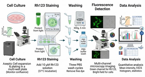 Fig. 2. Comprehensive workflow diagram of the Rhodamine 123 staining protocol (BOC Sciences Authorized).
1. Cell Preparation
- Cell culture and handling: First, select a suitable cell line for the experiment and culture it in an appropriate growth medium. To ensure reliable and reproducible results, maintain cell density within a reasonable range, typically at 50–70% confluence. The culture medium can be DMEM supplemented with 10% fetal bovine serum (FBS) or other media suitable for the specific cell line. During culture, cells should be incubated at 37°C in a 5% CO₂ incubator to maintain optimal growth conditions.
- Cell counting and seeding: Digest the cells with trypsin to detach them from the culture vessel. Count the cells using Trypan Blue staining to confirm cell viability. Seed the cells evenly in 96-well plates or other suitable culture plates, ensuring consistent cell numbers per well. After seeding, incubate the cells for 24 hours to allow them to adhere and recover normal physiological conditions.
2. Staining Procedure
- Preparation and dilution of Rh123 solution: Prepare Rh123 under sterile conditions. Dilute the stock solution to the desired working concentration, typically 5–10 µM, using pre-chilled calcium- and magnesium-free PBS to maintain dye stability.
- Detailed staining steps: Incubate cells with Rh123 at the selected working concentration (usually 5–10 µM) for 15–30 minutes. Incubation time affects fluorescence intensity and should be optimized for each experiment. Perform staining at 37°C to maintain normal membrane potential and mitochondrial function. After incubation, wash cells three times with pre-chilled PBS to remove unbound dye, then add fresh PBS for fluorescence detection.
3. Detection and Analysis
- Fluorescence microscopy: Observe stained cells using a fluorescence microscope equipped for Rh123 (excitation 488 nm, emission 530 nm). Capture images from several fields of view under identical exposure settings for all samples.
- Data acquisition and analysis: Use software like ImageJ to quantify fluorescence intensity in cells. Analyze differences between treatment groups and perform statistical analysis of results.
Precautions and Optimization Tips
Staining Precautions
- Avoid light-induced fluorescence loss: Store Rh123-stained samples away from light to prevent photobleaching. Perform procedures in a dark room or cover samples with foil.
- Ensure healthy cell status: Maintain cell integrity throughout staining and detection. Avoid over-digestion with trypsin, which can damage the cell membrane.
Common Issues and Solutions
- Insufficient or excessive staining:
- Insufficient staining may result from low dye concentration or short incubation. Increase Rh123 concentration or extend incubation time.
- Excessive staining may be caused by high dye concentration or prolonged incubation, leading to nonspecific staining. Reduce dye concentration or shorten incubation.
- Weak mitochondrial fluorescence: Weak fluorescence may indicate poor cell condition or impaired mitochondrial function. Ensure optimal culture conditions to enhance signal intensity.
Experimental Optimization Tips
- Optimize for different cell types: Sensitivity to Rh123 varies across cell lines; adjust concentration and incubation time accordingly. Cells with lower mitochondrial content may require higher dye concentrations and longer incubation.
- Improve reproducibility: Maintain consistent conditions across experiments, including cell density, dye concentration, incubation time, and temperature. Following standardized operating procedures (SOPs) enhances reproducibility and reliability.
Applications of Rhodamine 123 Staining
Rhodamine 123 has become an indispensable tool in cell biology and mitochondrial research due to its high specificity and strong fluorescence. It selectively accumulates in mitochondria, providing precise fluorescent signals that reflect mitochondrial membrane potential and cellular viability. The applications of Rh123 span fundamental scientific research, such as energy metabolism and apoptosis mechanism studies, as well as drug screening, toxicity assessment, and disease model research. With high sensitivity, low toxicity, and reproducibility, Rh123 offers researchers a reliable method to explore the complex relationship between cellular function and pathological processes.
Cell Viability Assessment
The intensity of Rh123 fluorescence directly reflects mitochondrial activity, allowing evaluation of overall cell viability. In healthy cells, normal mitochondrial membrane potential enables robust Rh123 accumulation, producing strong green fluorescence. In damaged or apoptotic cells, membrane potential decreases, dye accumulation is reduced, and fluorescence weakens. This highly sensitive detection method is suitable for routine monitoring under standard culture conditions and is widely used in toxicology assessments and physiological state analysis. For instance, in drug treatment or environmental stress experiments, changes in Rh123 signal intensity can quickly indicate cell health and potential toxicity, providing reliable metrics for experiment optimization and quality control.
Mitochondrial Function Analysis
Rh123 fluorescence intensity is positively correlated with mitochondrial membrane potential, making it a useful tool for quantitative assessment of mitochondrial function. Specific applications include monitoring mitochondrial depolarization, tracking dynamic changes in energy metabolism, and evaluating the impact of mitochondrial damage on cellular physiology. In basic research, Rh123 helps reveal the role of mitochondria in energy metabolism, oxidative stress responses, and signal transduction. In applied studies, it enables researchers to quantify the effects of drugs or genetic manipulations on mitochondrial function, providing deeper insights into the relationship between cellular metabolic state and physiological function.
Drug Screening and Toxicity Assessment
Rh123 is an important tool for studying drug mechanisms, particularly compounds affecting mitochondrial energy metabolism. By comparing Rh123 fluorescence before and after treatment, researchers can precisely quantify the extent of mitochondrial functional impact, evaluating drug efficacy and potential toxicity. For example, in anticancer drug development, Rh123 allows rapid screening of candidates that specifically target tumor cell mitochondria. In neurotoxicity testing, it enables assessment of compound-induced mitochondrial damage in neurons, providing quantitative data for safety evaluation.
Disease Model Research
In models of neurodegenerative diseases, myocardial ischemia/reperfusion injury, or metabolic disorders, Rh123 can visualize mitochondrial damage and reactive oxygen species (ROS) generation, aiding in the elucidation of disease mechanisms. For instance, in Parkinson's disease models, Rh123 can detect early changes in mitochondrial membrane potential. In cardiac ischemia models, it monitors mitochondrial functional recovery or damage extent. Furthermore, when combined with apoptosis markers or ROS probes, Rh123 provides clear, quantitative mitochondrial data for multiparametric disease model studies, supporting evidence-based evaluation of therapeutic interventions.
Fluorescent Dyes Recommended for Your Research Project
| Catalog | Name | CAS | Inquiry |
|---|---|---|---|
| A18-0008 | Rhodamine 110 chloride | 13558-31-1 | Bulk Inquiry |
| A14-0036 | Rhodamine B hydrazide | 74317-53-6 | Bulk Inquiry |
| A16-0093 | Rhodamine 6G | 989-38-8 | Bulk Inquiry |
| A17-0016 | Rhodamine 6G Perchlorate | 13161-28-9 | Bulk Inquiry |
| A17-0061 | Rhodamine 610 Perchlorate | 23857-51-4 | Bulk Inquiry |
| A16-0014 | Sulforhodamine 101 | 60311-02-6 | Bulk Inquiry |
| A01-0005 | Rhodamine B | 81-88-9 | Bulk Inquiry |
| A16-0142 | Dihydrorhodamine 6G | 217176-83-5 | Bulk Inquiry |
| A17-0069 | Rhodamine 590 Chloride | 3068-39-1 | Bulk Inquiry |
| A17-0062 | Rhodamine 3B Perchlorate | 23857-69-4 | Bulk Inquiry |
Corporate Profile
BOC Sciences is a leading independent provider of high-purity fluorescent chemicals, fluorescent dyes, and probes, offering comprehensive solutions for advanced cell imaging, mitochondrial assays, and fluorescent staining applications. With decades of experience in chemical supply and research services, BOC Sciences combines deep technical expertise with scientific innovation, supporting laboratories worldwide in achieving precise, reproducible, and reliable experimental results. Our mission is to empower researchers with high-quality reagents and professional guidance, enabling breakthroughs in cellular biology, molecular imaging, and pharmacological research.
BOC Sciences specializes in the development and supply of fluorescent probes, including Rhodamine 123, optimized for mitochondrial staining, cell viability analysis, and organelle-specific imaging. Each reagent undergoes rigorous quality control to ensure stability, high signal fidelity, and consistent performance under diverse experimental conditions. Beyond standard products, we offer custom synthesis, labeling, and conjugation services, allowing researchers to tailor probes to their specific experimental needs. This flexibility ensures optimal performance in complex assays, including studies of mitochondrial membrane potential, energy metabolism, and drug-induced cytotoxicity.
In addition to premium reagents, BOC Sciences provides extensive technical support and protocol guidance to maximize experimental outcomes. Our team assists researchers with staining procedure optimization, troubleshooting, and best practices for applying Rhodamine 123 and other fluorescent dyes in various cellular models. With stringent quality assurance and compliance measures, our products are reproducible, safe, and compatible with multiple cell types and imaging platforms. By combining top-quality reagents with expert guidance, BOC Sciences enables researchers to advance disease model studies, drug screening, and cellular functional analyses, reinforcing our position as a trusted partner in fluorescent staining and mitochondrial research.
Custom Fluorescent Solutions Designed for Your Experiments
- DNA StainingPrecise fluorescent dyes for clear DNA visualization and analysis.
- Lipid StainingFluorescent solutions for effective lipid structure imaging.
- Cell StainingAdvanced staining for detailed cell morphology and analysis.
- Protein StainingHigh-quality staining for accurate protein detection and imaging.
- Bacteria ImagingFluorescent solutions to visualize bacterial structures and activity.
- Cell ImagingVisualize and analyze live or fixed cells using advanced fluorescence.
- Molecular ImagingCutting-edge fluorescent solutions for deep molecular analysis.
- Fluorescence ImagingHigh-resolution imaging solutions for detailed fluorescence studies.
- BioconjugationCustom bioconjugation services for protein, peptide, and dye linking.
- Drug DeliveryTailored fluorescent solutions for efficient drug delivery research.
- Molecular DiagnosticsFluorescent probes and markers for precise molecular diagnostics.
- Flow CytometryFluorescent dyes and reagents for enhanced flow cytometry analysis.
High-Performance Fluorescent Tools for Your Research
- Nuclear Fluorescent Probes DNA-binding dyes for nucleus visualization.
- Mitochondrial Fluorescent Probes Targeted dyes for mitochondrial structure and function.
- Cell membrane Fluorescent Probes Surface-labeling dyes for membrane dynamics studies.
- Nitric Oxide (NO) & Reactive Oxygen Species (ROS) Probes for oxidative stress and signaling detection.
- Other Cell Fluorescent Probes Functional probes for diverse cellular imaging studies.
- Endoplasmic Reticulum Fluorescent Probes ER-targeted dyes for organelle structure analysis.
- pH Indicators Fluorescent sensors for intracellular pH monitoring.
- Fluorescent Probes Versatile tools for biomolecular and cellular imaging.
Explore More Topics
Online Inquiry
