

Rhodamine Staining Protocols: Tips for High-Quality and Reproducible Results
Rhodamine dyes are widely used labeling tools among researchers due to their high brightness, photostability, and excellent cell permeability. Whether in cell staining, tissue section labeling, pathogen detection, or mitochondrial function studies, Rhodamine staining can provide high-resolution and reproducible imaging results. This article delves into rhodamine staining protocols from principles, types, common methods, optimization strategies, to practical applications, offering researchers practical tips to enhance staining quality and experimental reproducibility.
What Is Rhodamine Stain?
Before performing staining experiments, understanding the definition, characteristics, and applicable scope of Rhodamine staining is a key step to optimize experimental conditions and achieve high-quality results. As a class of high-brightness fluorescent dyes, Rhodamine dyes have wide applications in visual labeling of cells and tissues. By clarifying their staining targets and comparative advantages over other fluorescent dyes, researchers can select appropriate experimental schemes more scientifically, improving staining reproducibility and reliability.
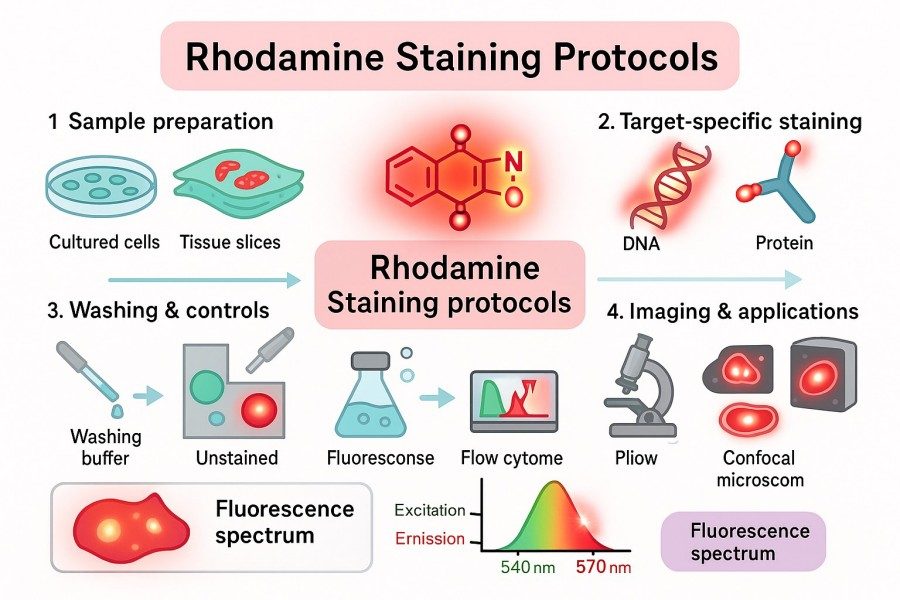 Fig. 1. Rhodamine staining protocols (BOC Sciences Authorized).
Rhodamine Stain Definition and Meaning
Rhodamine dyes are fluorescent compounds based on a xanthene (diphenylmethane) backbone, enabling strong red-orange fluorescence emission under UV or visible light excitation. Common representatives include Rhodamine B, Rhodamine 123, TRITC (Tetramethylrhodamine isothiocyanate), and Rhodamine 6G. These dyes not only exhibit high quantum yield and excellent photostability but can also bind specific targets in cells or tissues via covalent or non-covalent interactions, achieving high-contrast visualization. Thanks to these properties, Rhodamine dyes have become indispensable tools in cell imaging, protein localization, and histological analysis.
What Does Rhodamine Stain in Biological Samples?
Rhodamine dyes can stain a variety of biological structures specifically or semi-specifically, with broad applications in basic research and diagnostics. They can be used for cell and nucleus staining, allowing researchers to visualize cell morphology and nuclear structures. For the cytoskeleton, Rhodamine Phalloidin precisely labels actin filaments, enabling studies of cell migration and morphological changes. Rhodamine 123 is specialized for mitochondrial staining, accumulating in mitochondria in a membrane potential-dependent manner for functional monitoring. Additionally, the Auramine-Rhodamine staining method can detect bacteria and pathogens, particularly providing rapid fluorescent labeling of Mycobacterium tuberculosis, offering a reliable tool for pathogen research and clinical detection.
Advantages and Limitations Compared to Other Fluorescent Stains
Compared with other fluorescent dyes, Rhodamine dyes offer notable advantages. First, they provide high fluorescence brightness and excellent photostability, maintaining clear signals even during prolonged observation or repeated excitation. Second, Rhodamine exhibits good affinity for various cells and tissue structures, enabling multiplex staining experiments. Furthermore, it is compatible with multiple microscopy techniques, including confocal and fluorescence microscopy, ensuring stable and reliable imaging. However, Rhodamine dyes also have limitations: staining backgrounds can be high if washing is insufficient, some dyes may exert mild cytotoxicity requiring optimization of concentration and incubation time, and certain derivatives can experience fluorescence quenching in fixed samples, making the choice of appropriate anti-fade mounting media equally crucial.
Looking for Rhodamine Dyes?
From cell and tissue imaging to mitochondrial and pathogen analysis, our specialists provide tailored Rhodamine staining reagents and protocols to optimize your experimental results.
Rhodamine Staining Principles and Mechanisms
Understanding the principles and mechanisms of Rhodamine staining is key to ensuring controllable and reproducible results. This section explains how Rhodamine dyes bind to cellular and subcellular structures, their excitation and emission properties, and compatibility with microscopy and flow cytometry techniques, providing researchers with a scientific basis to optimize experimental conditions and reliability.
How Rhodamine Binds to Cellular and Subcellular Structures?
Rhodamine dyes target different cellular and subcellular structures, with binding mechanisms varying depending on the dye type. These mechanisms allow Rhodamine dyes to provide stable and specific labeling of cell membranes, nuclear structures, mitochondria, and the cytoskeleton.
- Non-covalent affinity: Rhodamine B can bind cell membranes or protein surfaces through electrostatic or hydrophobic interactions, achieving rapid staining.
- Covalent conjugation: TRITC (Tetramethylrhodamine isothiocyanate) forms stable covalent bonds with protein amino groups via its isothiocyanate group, enabling long-term labeling.
- Membrane potential accumulation: Rhodamine 123, a cationic dye, accumulates in mitochondria depending on the membrane potential for functional staining.
- High-specificity binding: Rhodamine Phalloidin binds F-actin, providing precise labeling of cytoskeletal actin filaments.
Excitation and Emission Characteristics of Rhodamine Dyes
The fluorescent properties of Rhodamine dyes make them highly effective imaging tools. Typically, Rhodamine dyes are excited in the 540–570 nm range and emit at 570–620 nm, producing bright red-orange fluorescence. High quantum yield ensures clear signals at low concentrations, while excellent photostability allows prolonged observation and repeated excitation without significant fading. These properties are particularly important for multiplex staining experiments, allowing combination with other fluorescent dyes, such as FITC or DAPI, for multicolor imaging without interference.
Compatibility with Microscopy and Flow Cytometry Techniques
Thanks to their multi-platform compatibility, researchers can flexibly choose detection methods based on experimental needs, obtaining reproducible, quantitative, and high-quality imaging results.
- Fluorescence Microscopy: Provides high-resolution imaging of cells and tissues, facilitating observation of cell morphology and subcellular structures.
- Laser Confocal Microscopy: Suitable for three-dimensional imaging and internal cellular structure analysis, with stable and reproducible signals.
- Flow Cytometry: Enables quantitative analysis of intracellular Rhodamine signals, applicable for functional studies and statistical evaluation.
- High-Throughput Imaging Platforms: Ideal for drug screening and large-scale cell function assessment, ensuring stable and reliable fluorescence signals.
Common Types of Rhodamine Staining
Rhodamine dyes are widely used in cell and tissue labeling due to their high brightness and target specificity. Different Rhodamine derivatives can precisely stain specific structures such as cell membranes, mitochondria, cytoskeletons, and pathogens. Understanding the various types of Rhodamine staining and their characteristics helps researchers select the most appropriate dye and method, achieving high-quality and reproducible results in imaging, functional analysis, and pathology studies.
Auramine Rhodamine Stain for Bacteria Detection
The Auramine-Rhodamine staining method is a classic technique for bacterial detection, widely applied in pathogen research and clinical diagnostics. This method combines Auramine with Rhodamine to enhance the fluorescent signal, causing bacteria to appear bright red or orange-red under a microscope. Particularly for Mycobacterium tuberculosis detection, Auramine-Rhodamine staining offers high sensitivity and rapid visualization, enabling accurate localization even in low bacterial load samples. Researchers can optimize staining time and washing steps to reduce background signals and improve detection reliability.
Tip: For low bacterial load samples, extending the staining incubation and increasing washing cycles can effectively reduce non-specific background and improve the signal-to-noise ratio.
Rhodamine 123 Staining for Mitochondria
Rhodamine 123 is a cationic fluorescent dye that specifically targets mitochondria, accumulating within them in a membrane potential-dependent manner for functional staining. It is commonly used to assess mitochondrial activity, cellular energy metabolism, and apoptosis. As the dye is light-sensitive, prolonged exposure may lead to fluorescence quenching, so experiments should be conducted under minimal light, with timely imaging.
Tip: The concentration and incubation time of Rhodamine 123 should be optimized according to cell type to avoid excessive accumulation that could interfere with mitochondrial function.
Rhodamine Phalloidin Staining for Actin Filaments
Rhodamine Phalloidin, a conjugate of Phalloidin and Rhodamine, binds F-actin with high specificity, labeling cytoskeletal actin filaments. This staining method is widely used in cell morphology studies, migration analysis, and signaling pathway research. Compared with other staining techniques, Rhodamine Phalloidin provides clear images of the cytoskeleton, aiding researchers in analyzing dynamic cell changes and tissue structures.
Tip: Staining immediately after fixation and permeabilization yields the best actin signal while minimizing non-specific background.
Rhodamine Cell and Nucleus Staining
Rhodamine dyes can also stain cell membranes and nuclei, providing clear contrast of cellular structures. Often combined with blue dyes such as DAPI or Hoechst, they enable multicolor fluorescence imaging, facilitating simultaneous observation of nuclear structures and cell morphology. In tissue sections or cultured cell experiments, this staining method helps quickly assess cell health and locate specific cell populations.
Tip: Selecting an appropriate Rhodamine dye concentration and optimizing incubation time helps reduce cytotoxicity and background signals.
Rhodamine B Staining and Its Applications
Rhodamine B is a versatile fluorescent dye suitable for various tissue staining and material labeling experiments. Its chemical stability and high brightness allow long-term signal retention in fixed samples, making it ideal for histological observation and extended imaging studies. Rhodamine B can be used for cell membranes, tissue sections, and biomaterial labeling, serving as a common fluorescent tool in basic and applied research.
Tip: For fixed tissue samples, using anti-fade mounting media can prolong fluorescence stability and improve experimental reproducibility.
Rhodamine Staining Protocols: Best Practices and Optimization
Mastering best practices and optimization strategies is key to achieving high-quality and reproducible results in Rhodamine staining experiments. Staining outcomes are influenced not only by dye type but also by sample preparation, staining conditions, solution preparation, and washing steps. Through scientific design and operational optimization, researchers can significantly improve signal-to-noise ratio, reduce non-specific background, and extend fluorescence stability, ensuring reliable imaging and analysis results.
Standard Rhodamine Staining Protocol for Cell and Tissue Samples
A standard Rhodamine staining workflow generally includes sample fixation, permeabilization, dye incubation, washing, and mounting for imaging:
- Fixation: Use 4% paraformaldehyde or other suitable fixatives to preserve cell or tissue structure.
- Permeabilization: For intracellular target staining, use Triton X-100 or Tween-20 to enhance dye penetration.
- Staining: Select an appropriate Rhodamine derivative and optimize concentration and incubation time.
- Washing: Thoroughly wash samples to remove non-specifically bound dye and reduce background fluorescence.
- Mounting & Imaging: Use anti-fade mounting media to maintain fluorescence stability and image with a fluorescence or confocal microscope.
Choosing the Right Rhodamine Derivative (e.g., Rhodamine B, TRITC, Rhodamine 6G)
Different Rhodamine derivatives target different structures and experimental requirements:
- Rhodamine B: A general-purpose dye suitable for cell membrane and tissue section staining.
- TRITC: Forms covalent bonds with proteins via its isothiocyanate group, suitable for long-term protein labeling.
- Rhodamine 123: Targets mitochondria for functional studies and membrane potential analysis.
- Rhodamine 6G: Ideal for rapid labeling and microbial detection, offering high fluorescence brightness.
Choosing the right dye ensures high-specificity labeling and avoids spectral interference, enabling multicolor co-staining.
Rhodamine Staining Solution Preparation and Storage
Proper preparation and storage of staining solutions directly affect experimental outcomes:
- Dissolution: Dissolve in PBS or an appropriate buffer according to dye properties, ensuring thorough mixing.
- Concentration: Optimize based on cell type and experimental purpose to balance signal strength and minimize cytotoxicity.
- Storage: Store protected from light at 4 °C. For short-term experiments, prepare fresh solutions; for long-term storage, aliquot and avoid repeated freeze-thaw cycles.
Strategies to Minimize Non-Specific Binding and Background Fluorescence
To achieve high-quality staining images, the following optimization strategies are recommended:
- Block non-specific sites: Pre-treat samples with BSA, normal serum, or other blocking agents.
- Optimize staining time and concentration: Excessive concentration or prolonged incubation may cause background signals; adjust based on cell type.
- Thorough washing: Perform multiple buffer washes after staining to remove residual unbound dye.
- Use anti-fade mounting media: Mounting enhances fluorescence stability, especially for prolonged imaging or repeated excitation experiments.
By applying these optimization measures, researchers can effectively improve the specificity and reproducibility of Rhodamine staining, providing reliable data for cell and tissue imaging.
Rhodamine Staining in Research and Diagnostics
Rhodamine staining plays a key role not only in basic cell and tissue research but also in pathogen detection, mitochondrial functional analysis, and cytoskeletal studies. In both research and clinical diagnostics, selecting the appropriate Rhodamine dye and staining strategy can significantly enhance signal specificity and experimental reproducibility, providing reliable tools for assessing cell function and conducting pathological analysis.
Rhodamine Staining of Liver Tissue (Wilson's Disease and Other Applications)
In liver tissue research, Rhodamine dyes can specifically label metal deposits, such as copper ions, making them useful for studies of Wilson's disease and other hepatic metabolic disorders. When working with thick tissue sections, researchers may extend dye penetration time and ensure thorough washing to reduce non-specific signals and obtain clear images of copper distribution. Additionally, Rhodamine staining can be applied in experimental models of liver fibrosis or steatosis to observe changes in cell morphology and tissue structure, providing reliable reference for pathological analysis.
Rhodamine Staining in Bacteria and Pathogen Studies
Auramine-Rhodamine staining is a commonly used fluorescence-based method in microbiology, particularly suited for rapid detection of Mycobacterium tuberculosis and other bacteria. By enhancing red fluorescence signals, this method allows easy visualization even at low bacterial concentrations. For low-load samples, researchers can extend staining incubation and perform multiple washes to improve the signal-to-noise ratio and detection accuracy. Moreover, Rhodamine staining can be combined with immunolabeling or molecular probes to achieve multiplex labeling and functional analysis, providing highly sensitive tools for pathogen research.
Rhodamine Staining of Mitochondria for Functional Studies
Rhodamine 123 is the preferred dye for studying mitochondrial function, accumulating within mitochondria in a membrane potential-dependent manner to reflect mitochondrial activity and metabolic status. In drug screening, apoptosis research, and cellular energy metabolism analysis, Rhodamine 123 staining provides direct mitochondrial signals suitable for quantitative analysis and dynamic monitoring. During experiments, prolonged light exposure and high dye concentrations should be avoided to prevent mitochondrial damage or fluorescence quenching, ensuring reliable staining results.
Rhodamine Actin Staining in Cytoskeletal Analysis
Rhodamine Phalloidin is a widely used tool for analyzing the cytoskeleton and actin filaments. By binding F-actin with high specificity, this staining method enables observation of cell morphology, migration behavior, and signaling pathways. In tissue section or cultured cell experiments, researchers should stain immediately after fixation and permeabilization and avoid extended incubation to maintain actin structure integrity and clear fluorescence signals, yielding high-quality cytoskeletal imaging data.
Rhodamine Stain Copper and Other Special Stains
Beyond conventional cell and tissue labeling, Rhodamine dyes can be used for specific element and molecular detection, such as copper ions, calcium ions, and certain drug targets. These applications often utilize fluorescence microscopy or multiphoton imaging to analyze the spatial distribution of particular molecules or metals. For metal or drug staining, researchers should use low-salt buffers and perform procedures in the dark to preserve fluorescence stability and sample integrity, achieving high-resolution quantitative data.
Key Considerations for Successful Rhodamine Staining
To ensure high-quality and reproducible Rhodamine staining results, researchers should consider multiple factors, including dye selection, operational conditions, experimental design, and staining limitations.
Choosing the Right Rhodamine Dye for Your Research
Different Rhodamine derivatives are suited for distinct experimental purposes and targets. When selecting a dye, researchers should consider sample type, target structure, and imaging technique. For example, Rhodamine 123 specifically targets mitochondria for energy metabolism and functional studies; Rhodamine Phalloidin binds F-actin with high specificity for cytoskeletal analysis; TRITC enables long-term protein labeling via covalent binding. Fluorescence spectra compatibility with other dyes should also be considered for multicolor co-staining without spectral interference.
Avoiding Common Pitfalls in Rhodamine Staining Protocols
Common issues include excessive non-specific background, fluorescence signal decay, and potential dye toxicity to cells or tissues. To prevent these problems, researchers should optimize dye concentration and incubation time according to sample type, strictly control washing steps to remove unbound dye, and minimize light exposure to prevent fluorescence quenching. For fixed samples, choosing suitable mounting media and anti-fade agents is crucial to extend signal stability and reproducibility.
Rhodamine Stain Purpose and Limitations
Although Rhodamine dyes are widely used in research and diagnostics, they have limitations. Certain derivatives may experience fluorescence quenching under prolonged imaging or high-intensity light. At specific concentrations, they may exhibit mild cytotoxicity, and staining background can be relatively high. Therefore, experimental design should clearly define staining goals, carefully select dye type, concentration, and incubation time, and optimize conditions to balance signal intensity, specificity, and cell or tissue health.
How BOC Sciences Supports Your Research with Rhodamine Staining?
In modern biological research and drug development, high-quality, reproducible fluorescent staining is key for successful cell imaging, protein localization, mitochondrial functional analysis, and pathogen detection. BOC Sciences, as a leading supplier of fluorescent products, provides not only a wide range of high-quality Rhodamine dyes but also customized staining services and technical support. We help researchers, biotech companies, and pharmaceutical firms optimize experimental workflows, enhance staining specificity, and maintain signal stability. Leveraging extensive experimental experience and strict quality control, BOC Sciences delivers complete solutions for diverse research and application scenarios.
Rhodamine Staining for Cell Imaging
- Offers multiple Rhodamine dyes for high-resolution imaging of cell membranes, nuclei, and cytoskeletons.
- Optimizes dye concentration, incubation time, and washing steps for different cell types to ensure signal strength and specificity.
- Provides detailed protocols and technical support to reduce non-specific background and signal decay.
- Supports multicolor co-staining experiments, compatible with common dyes like FITC and DAPI for complex imaging setups.
Rhodamine Staining for Protein Localization
- Supplies TRITC, Rhodamine B, and other dyes for precise protein labeling and long-term tracking studies.
- Optimizes staining protocols for fixed and live cells to ensure accurate and reproducible protein localization.
- Supports multicolor co-staining techniques to avoid spectral interference and improve data accuracy and comparability.
- Offers technical consulting and optimization guidance for analyzing protein interactions and signaling pathways.
Rhodamine Staining for Mitochondrial Research
- Provides mitochondrial-specific dyes such as Rhodamine 123 for membrane potential and metabolic function analysis.
- Optimizes dye concentration and light exposure conditions for drug screening and apoptosis studies to maintain signal stability.
- Supports high-resolution confocal microscopy imaging for dynamic observation and quantitative analysis of mitochondria.
- Enables live-cell staining experiments to preserve cell function and extend fluorescence signal stability.
Rhodamine Staining for Microbial Detection
- Offers Auramine-Rhodamine staining protocols for highly sensitive detection of Mycobacterium tuberculosis and other pathogens.
- Optimizes staining and washing conditions based on sample type to ensure accurate detection in low bacterial load samples.
- Supports multiplex labeling experiments, combining with immunostaining or molecular probes for high-precision localization analysis.
- Provides operational guidance and experimental optimization to improve reproducibility and reliability of detection results.
Do You Need A Consultation?
BOC Sciences integrates cutting-edge fluorescence technologies to accelerate your research, driving next-generation solutions for drug discovery and diagnostics.
Transform Your Studies with Cutting-Edge Fluorescent Products
| Catalog | Name | CAS | Inquiry |
|---|---|---|---|
| A19-0011 | 5-Carboxytetramethylrhodamine succinimidyl ester | 150810-68-7 | Bulk Inquiry |
| F07-0049 | 6-Carboxytetramethylrhodamine succinimidyl ester | 150810-69-8 | Bulk Inquiry |
| A19-0008 | Tetramethylrhodamine-5-Iodoacetamide Dihydroiodide | 114458-99-0 | Bulk Inquiry |
| A17-0047 | Rhodamine 700 perchlorate | 63561-42-2 | Bulk Inquiry |
| A03-0012 | Dihydrorhodamine 123 | 109244-58-8 | Bulk Inquiry |
| A16-0054 | Tetramethylrhodamine ethyl ester perchlorate | 115532-52-0 | Bulk Inquiry |
| A16-0053 | Tetramethylrhodamine methyl ester perchlorate | 115532-50-8 | Bulk Inquiry |
| A16-0019 | Tetramethylrhodamine isothiocyanate (mixed isomers) | 95197-95-8 | Bulk Inquiry |
| F07-0023 | 5-(6)-Carboxytetramethylrhodamine | 98181-63-6 | Bulk Inquiry |
| F05-0008 | 5-Carboxy-x-rhodamine triethylammonium salt | 216699-35-3 | Bulk Inquiry |
High-Performance Fluorescent Tools for Your Research
- Cyanine3 Standard green-orange fluorescent biomolecular labeling.
- ICG Dyes Near-infrared imaging and in vivo diagnostics.
- BODIPY Photostable dyes for lipid and cell imaging.
- sulfo-Cyanine5 Hydrophilic red fluorescent bioconjugation.
- Fluorescent Dyes General-purpose labeling for bioanalytical detection.
- Cyanine7.5 Extended NIR imaging for in vivo studies.
- Fluorescein FAM Green fluorescence for nucleic acid labeling.
- sulfo-Cyanine3.5 Water-soluble orange-red fluorescent labeling.
Explore More Topics
Online Inquiry
