

Lipid Staining with BODIPY Dyes: Accurate Visualization of Lipid Structures
In cellular lipid research, accurately locating and tracking lipid droplets, membrane structures, and lipid metabolic pathways is crucial. BODIPY dyes have become ideal tools for lipid staining due to their strong lipophilicity, high fluorescence intensity, and excellent photostability. They are used in various applications such as lipid droplet imaging in live cells, fatty acid uptake analysis, and membrane structure labeling. However, challenges remain, including low signal-to-noise ratio in dense lipid environments and photobleaching of traditional dyes. This article systematically explores how BODIPY dyes overcome these technical bottlenecks and demonstrates their broad application and customization potential in lipid visualization.
Introduction to Lipid Staining in Biological Research
Lipids are fundamental components of cellular life, widely distributed in biological membranes, energy storage particles, signaling molecules, and metabolic pathways. In cell biology, metabolic studies, drug screening, and disease mechanism exploration, accurate visualization of lipid distribution and dynamic changes has become a major focus. To achieve clear observation of lipid structures, fluorescent staining has become the preferred tool among researchers. Among these tools, BODIPY (boron-dipyrromethene) dyes are gradually replacing traditional dyes due to their excellent fluorescence properties, lipophilicity, and chemical tunability, becoming key reagents in lipid staining.
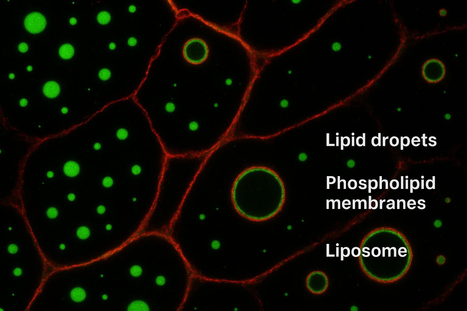
Fig. 1. Lipid staining in biological research (BOC Sciences Authorized).
Why Lipid Staining Matters in Cell Biology and Metabolism?
Lipids are not only the structural basis of cell membranes but also play key roles in signal transduction, energy storage and release, organ development, apoptosis, and inflammatory responses. For example, lipid droplets are critical organelles for storing neutral lipids and directly reflect the metabolic state of cells through their dynamic changes. The composition and integrity of phospholipid membranes are closely related to cell homeostasis, apoptosis, and membrane protein function. Abnormal lipid metabolism has become a central concern in the study of metabolic diseases, cancer, and neurodegenerative disorders. Therefore, building a precise, high-resolution lipid imaging system can not only aid basic research but also support drug development and early disease diagnosis.
Key Lipid Structures Commonly Visualized
In lipid staining, different lipid structures exhibit varying selectivity and affinity toward fluorescent probes, necessitating targeted dye selection. The three most commonly analyzed structures are:
- Lipid Droplets (LDs): Organelles storing triglycerides and cholesterol esters, found widely in hepatocytes, adipocytes, and tumor cells. They are crucial in studies of energy metabolism and lipotoxicity.
- Phospholipid Membranes: Composed mainly of phospholipids and cholesterol, they maintain cell structure and regulate transport, serving as platforms for signal molecule aggregation.
- Liposomes: Artificial phospholipid bilayer vesicles used in drug delivery and biomembrane simulation; they are hot research tools in pharmacology and material science.
Techniques for Detecting Neutral and Polar Lipids
Modern techniques such as confocal microscopy, flow cytometry, and high-throughput screening, combined with fluorescent probes, allow multidimensional monitoring of lipid types, distribution, quantity, and dynamics. Lipid staining must consider lipid polarity:
- Neutral lipids (e.g., triglycerides, cholesterol esters) tend to aggregate in lipid droplets and are suitable for staining with strongly hydrophobic dyes such as BODIPY 493/503.
- Polar lipids (e.g., phospholipids, sphingolipids) are mainly distributed in cell or organelle membranes and require dyes with some hydrophilicity or membrane-insertion capability.
Challenges in Lipid Visualization Workflows
Despite notable progress in lipid staining—especially in fluorescent probe development and imaging platform upgrades—researchers still face several technical challenges in practice. These issues not only affect imaging quality and analytical accuracy but also compromise the feasibility and reproducibility of experimental designs.
Low Signal-to-Noise Ratio in Dense Lipid Environments
In some cell types or pathological conditions, large amounts of dense lipid droplets or other lipid aggregates may form. These high-density environments lead to signal overlap and background interference. Fluorescent probes may generate overly strong local signals after entering lipid droplets, obscuring details of surrounding structures. Moreover, the heterogeneous lipid distribution causes significant signal intensity variation, lowering overall image signal-to-noise ratio, and hindering accurate assessment of lipid quantity, size, and spatial localization. This limits the quantitative analysis of lipid metabolism dynamics.
Photobleaching and Limited Stability of Traditional Dyes
Traditional lipid dyes such as Nile Red and Oil Red O undergo rapid photobleaching under prolonged excitation light, leading to fast fluorescence decay. This makes them unsuitable for long exposures, time-lapse acquisition, or real-time tracking. Additionally, they exhibit poor stability under light and during storage, prone to quenching, drift, or spectral shifts—affecting experimental reproducibility and data comparability.
Inadequate Specificity for Subtypes of Lipids
Lipids encompass a wide variety, including neutral lipids (e.g., triglycerides, cholesterol esters) and polar lipids (e.g., phospholipids, sphingolipids). Many traditional dyes lack sufficient structural specificity and tend to label all lipids non-selectively. For example, some dyes can enter both lipid droplets and embed into membranes, producing mixed fluorescence patterns that complicate lipid localization, subtype quantification, and functional analysis in images.
Compatibility Issues with Live vs. Fixed Cell Staining
Lipid research requires both high-resolution imaging in fixed cells and dynamic monitoring in live cells. However, not all dyes are compatible across platforms. Some work only in fixed cells and perform poorly in live cells due to poor uptake, hydrolysis, membrane impermeability, or high cytotoxicity. Some dyes may even disrupt normal lipid metabolism, causing experimental bias. This lack of adaptability severely limits their use in long-term imaging, time-lapse tracking, or in vivo studies.
Why Use BODIPY Dyes for Lipid Staining?
BODIPY dyes have become essential tools in lipid visualization due to their outstanding optical properties, stable chemical structures, and high tunability. Compared to traditional dyes, BODIPY offers significant advantages in lipophilicity, fluorescence intensity, photostability, and biocompatibility. These features make them especially suitable for demanding real-time imaging, multiplex labeling, and live-cell staining—greatly enhancing the resolution and reliability of lipid studies.
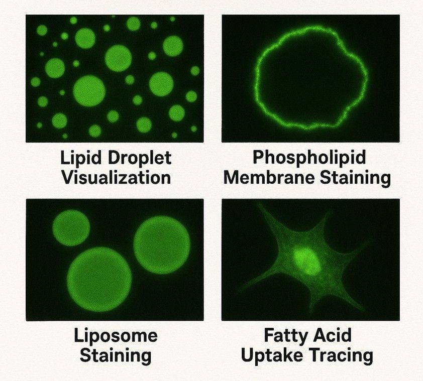
Fig. 2. BODIPY dyes for lipid staining (BOC Sciences Authorized).

Lipophilic Nature Enables High Affinity for Lipid Structures
The molecular structure of BODIPY dyes contains highly hydrophobic groups, providing strong lipophilicity that allows stable insertion into lipid droplets, neutral lipids, or phospholipid bilayers. This results in superior accumulation within lipid structures compared to conventional dyes, enhancing staining specificity and signal intensity for structures such as lipid droplets, liposomes, and membranes.

Strong Fluorescence, High Photostability, and Narrow Spectra
BODIPY dyes exhibit excellent quantum yields and stable luminescence, delivering strong, clear signals even at very low concentrations. Their photobleaching rate is much lower than that of traditional dyes, enabling sustained brightness during long exposures or laser scanning. Furthermore, their narrow emission spectra reduce signal crosstalk, improving resolution in multicolor imaging.

Tunable Emission Across Visible and NIR Range for Multiplexing
BODIPY dyes have highly tunable structures. By introducing different substituents, their emission wavelengths can be adjusted from the visible to the near-infrared (NIR) region. This allows them to be designed as green, red, far-red, or even NIR channel dyes, making them compatible with other fluorescent tags and well-suited for multiplex and multi-channel microscopy.

Low Cytotoxicity and Suitability for Live Cell Imaging
Compared to some traditional lipid dyes, BODIPY dyes provide chemical stability and lipophilicity that support effective staining without disturbing membrane integrity or lipid metabolism. They exhibit low cytotoxicity and are suitable for live-cell, tissue section, and even small animal in vivo imaging. This enables high-quality dynamic lipid visualization while maintaining cell viability.
BODIPY Dyes at BOC Sciences
| Cat. No. | Product Name | CAS No. | Inquiry |
|---|---|---|---|
| F01-0166 | BODIPY 493/503 NHS Ester | 216961-98-7 | Inquiry |
| F01-0185 | BODIPY FL C12 | N/A | Inquiry |
| F01-0186 | BODIPY FL C5 | N/A | Inquiry |
| R12-0001 | BODIPY 493/503 | 121207-31-6 | Inquiry |
| F01-0044 | BODIPY-Cholesterol | 878557-19-8 | Inquiry |
| F01-0177 | BODIPY FL Hydrazide | N/A | Inquiry |
| F01-0154 | BODIPY FL acid | 126250-45-1 | Inquiry |
| F01-0161 | BODIPY 558/568 C12 | 158757-84-7 | Inquiry |
Applications of BODIPY Dyes in Lipid Staining
Whether in studies of metabolism-related diseases, analyses of cell membrane structures, or investigations of liposome-based drug delivery systems, BODIPY dyes enable highly sensitive, highly specific, and high-resolution lipid labeling. Their excellent photostability and low toxicity also make them particularly outstanding in real-time live-cell imaging. The following are several typical applications of BODIPY dyes in lipid staining.
Lipid Droplet Visualization in Metabolism Studies
Lipid droplets are essential organelles for storing neutral lipids within cells and are widely found in metabolically active tissues such as hepatocytes, adipocytes, and muscle cells. For example, BODIPY 493/503 is one of the most commonly used dyes for lipid droplets. With strong lipophilicity, it can precisely embed into the core of lipid droplets and emit green fluorescence with a high signal-to-noise ratio. Researchers use this dye to evaluate lipid accumulation levels under conditions such as nutrient deprivation, insulin stimulation, or fatty acid overload, supporting the study of the pathogenesis of metabolic syndrome, fatty liver, obesity, and type II diabetes.
Staining of Phospholipid Membranes and Liposomes
Phospholipid membranes are the fundamental structural units of cells and key platforms for many membrane proteins, signaling pathways, and drug transport mechanisms. BODIPY dyes like BODIPY FL C5-HPC, which contain phospholipid tail groups, can embed into phospholipid bilayers and emit bright fluorescence. They are commonly used to label cell membranes, endoplasmic reticulum membranes, or artificially prepared liposomes. These dyes have great application value in studies of transmembrane protein transport, membrane fusion processes, and simulated nanocarrier systems, especially suitable for membrane biophysics experiments and visual tracking of drug release behavior.
Tracing Fatty Acid Uptake and β-Oxidation
BODIPY-C12 is a probe modified with a BODIPY group at the fatty acid tail, capable of mimicking the behavior of natural long-chain fatty acids. After entering the cell, it can be endocytosed or transported via carriers and then participate in mitochondrial β-oxidation. Through real-time imaging, researchers can observe the intracellular transport and metabolic conversion of fatty acids, quantitatively assessing the uptake rate and metabolic activity under different treatment conditions. This holds significant value in studies of metabolic diseases, lipotoxicity, liver dysfunction, and nutrition.
Monitoring Lipid Dynamics in Real-Time Live Imaging
Due to its strong fluorescence signal, extremely low photobleaching rate, and excellent cell compatibility, BODIPY dyes are especially suitable for long-term observation of lipid structures in live cells. Researchers can use confocal microscopy or live-cell imaging systems to track the formation, fusion, degradation, and intracellular transport of lipid droplets, revealing the dynamic mechanisms of lipid homeostasis regulation. This type of dynamic imaging plays an irreplaceable role in studies of energy metabolism regulation, cellular stress responses, lipid signaling pathways, and early warnings of lipid metabolism disorders.
Custom BODIPY Fluorescent Probe Services
With the continuous advancement of lipid visualization technologies, standard dyes can no longer meet the complex experimental demands for imaging precision, lipid type selectivity, and platform compatibility. Our customized probe services based on BODIPY dyes are providing more efficient and reliable technical support for lipid research, drug development, and in vivo imaging.
Design of Tailored BODIPY Structures for Specific Lipid Targets
We offer fine-tuned modifications of the BODIPY core based on the target lipid types (e.g., sphingolipids, phosphatidylinositol, cholesterol esters), introducing recognition groups or adjusting hydrophobicity to achieve selective staining of specific lipid components, thereby enhancing signal specificity and imaging clarity.Conjugation with Fatty Acids, Cholesterol, and Other Lipid Moieties
Through chemical conjugation, the BODIPY structure can be linked to lipid molecules such as fatty acids, cholesterol, and ceramides, creating functional probes. For example, BODIPY-C12 is used to trace fatty acid metabolic pathways, while BODIPY-Chol is used to observe the distribution and movement of cholesterol in cell membranes and the endoplasmic reticulum.Optimization for Flow Cytometry, Confocal Imaging, or HTS
We can optimize the probe's spectral properties, enhance its stability, or improve channel compatibility based on different experimental platforms (e.g., FACS flow cytometry, CLSM confocal imaging, SIM/STED super-resolution imaging, HTS high-throughput screening) to ensure signal strength and operational adaptability, thereby improving experimental efficiency.Scalable Production with Batch Consistency and QC Support
In addition to laboratory-scale customization, we also offer pilot and industrial-scale batch synthesis services, suitable for large research projects, diagnostic development, and drug screening platforms. All product batches can be provided with detailed quality control reports to ensure consistent performance and reliability across experimental batches.
Do You Need A Consultation?
BOC Sciences integrates cutting-edge fluorescence technologies to accelerate your research, driving next-generation solutions for drug discovery and diagnostics.
Versatile Fluorophores for Modern Labs
| Cat. No. | Product Name | CAS No. | Inquiry |
|---|---|---|---|
| F01-0155 | BODIPY Fl C5-Ceramide | 133867-53-5 | Inquiry |
| F01-0184 | BODIPY 558/568 C12, SE | N/A | Inquiry |
| F01-0158 | BODIPY TR methyl ester | 150152-63-9 | Inquiry |
| F01-0162 | BODIPY FL Prazosin | 175799-93-6 | Inquiry |
| F01-0189 | BODIPY FL-C5 NHS Ester | N/A | Inquiry |
| F01-0195 | BODIPY FL C5-Sphingomyelin | N/A | Inquiry |
| F01-0046 | Bodipy C12-Ceramide | 1246355-58-7 | Inquiry |
| F01-0203 | BODIPY FL Cholesterol | N/A | Inquiry |
| F01-0041 | Olaparib-bodipy FL | 1380359-84-1 | Inquiry |
| F01-0254 | BODIPY 493/503 carboxylic acid | 216961-95-4 | Inquiry |
High-Performance Fluorescent Tools for Your Research
- Fluorescent Dyes General-purpose labeling for bioanalytical detection.
- Other Cyanine Tunable near-infrared bioimaging and labeling.
- sulfo-Cyanine Water-soluble cyanine dyes for labeling.
- Alexa Fluor Bright, photostable dyes for fluorescence imaging.
- TAMRA Dyes Red fluorescent labeling for biomolecule tracking.
- sulfo-Cyanine3 Hydrophilic green-orange fluorescent conjugation.
- sulfo-Cyanine5 Hydrophilic red fluorescent bioconjugation.
- Cyanine Versatile fluorophores for bioimaging applications.
Explore More Topics
Online Inquiry
