

BODIPY Dyes for High-Performance Flow Cytometry Analysis
In today's era of flow cytometry emphasizing high sensitivity and multiparameter analysis, the selection of fluorescent dyes has become a critical factor determining experimental success. BODIPY (boron-dipyrromethene) dyes, owing to their unique structure and excellent optical properties, are gradually becoming ideal tools for high-performance flow cytometry analysis. Compared to traditional fluorescent dyes, BODIPY exhibits significant advantages such as tunable emission wavelengths, high fluorescence quantum yield, strong photostability, and low background signal. These characteristics make it particularly suitable for complex multicolor panel designs and long-term acquisition experiments.
What is Flow Cytometry?
Flow cytometry is a powerful biomedical analytical technology that can quickly and accurately measure a variety of biophysical and biochemical characteristics of cells at the single-cell level. Its basic principle involves illuminating a stream of cell suspension with lasers and detecting the scattered light and fluorescence signals emitted by the cells to perform quantitative analysis and sorting. Flow cytometry dates back to the 1960s and has become a core tool in cell biology, immunology, oncology, and hematology with continuous technological advances.
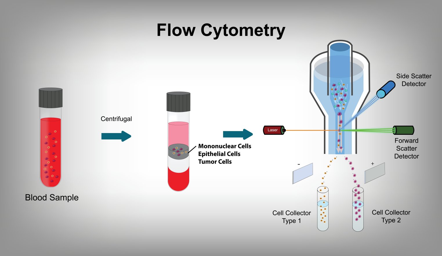
Fig. 1. Flow cytometry (BOC Sciences Authorized).
The core components of a flow cytometer include a flow chamber, laser light source, optical system, and data acquisition system. The cell suspension flows through the laser focus in the flow chamber in a single-cell fashion. After interaction with the laser, the cells produce scattered and fluorescent light. These signals are collected and spectrally separated by the optical system and then converted into electrical signals by detectors such as photomultiplier tubes or photodiodes. These signals are subsequently processed by the data acquisition system to generate analyzable flow cytometry data.
Key Applications of Flow Cytometry
Flow cytometry has wide applications in biomedical research, covering areas such as cell phenotype analysis, cell cycle detection, apoptosis studies, cell sorting, and functional analysis.
- Cell Phenotype Analysis: By labeling cell surface markers, flow cytometry can rapidly distinguish between different cell types, such as identifying T cells, B cells, and monocytes in immunology.
- Cell Cycle Detection: By using dyes (e.g., PI or DAPI) to stain DNA and measure DNA content changes, flow cytometry can analyze different stages of the cell cycle (G0/G1, S, G2/M).
- Apoptosis Studies: Flow cytometry can monitor apoptosis through detection of phosphatidylserine externalization (Annexin V labeling) or DNA fragmentation (TUNEL assay).
- Cell Sorting: Flow cytometry not only analyzes cells but can also sort target cells based on specific fluorescence signals for further experimental research or clinical applications.
- Functional Analysis: For example, detecting intracellular reactive oxygen species or calcium ion concentration changes provides important tools for studying cell signaling and physiological functions.
Challenges in Flow Cytometry-Based Research
Despite its powerful capabilities, flow cytometry still faces several technical challenges in practice, particularly regarding the selection and application of fluorescent dyes.
- Spectral Overlap in Multiparameter Analysis: In multiplex experiments, emission spectra of different fluorescent probes may significantly overlap, causing signal crosstalk and reducing data accuracy. This spectral overlap limits the number of parameters that can be analyzed simultaneously and increases the complexity of compensation calculations.
- Photobleaching and Signal Degradation: Many traditional dyes suffer from poor photostability and easily bleach under continuous excitation, leading to rapid signal loss, especially during long-term detection or imaging.
- Low Sensitivity in Rare Cell Detection: When the target cells constitute a very small proportion of the total population (<0.1%), probes with insufficient signal intensity may fail to distinguish positives from background noise, affecting accurate rare event detection.
- Difficulties in Labeling Small Molecules or Lipids: Conventional fluorescent dyes often exhibit low binding efficiency, high non-specific background, and weak signals when labeling small molecules or hydrophobic lipids, impacting subsequent data analysis.
Fluorescent Probes for Flow Cytometry
Fluorescent probes are one of the core tools in flow cytometry, and their performance directly affects the accuracy and reliability of experimental results. Selecting the appropriate fluorescent dye is critical for successful experiments.
Importance of Choosing the Right Fluorophore
Selecting a fluorescent dye requires consideration of multiple factors, including its spectral properties, chemical stability, fluorescence intensity, photobleaching characteristics, and binding capability to biomolecules. An ideal dye should possess a narrow emission spectrum, high quantum yield, good photostability, and the ability to specifically bind to target molecules without affecting their biological functions.
Comparison of Common Fluorescent Dyes
Common fluorescent dyes include FITC, PE, PerCP, and APC. Each of these dyes has its own advantages and disadvantages.
| Fluorescent Dye | Excitation Wavelength (nm) | Emission Wavelength (nm) | Brightness | Photostability | Advantages | Disadvantages | Recommended Applications |
|---|---|---|---|---|---|---|---|
| FITC | 488 | 519 | Moderate | Poor | Low cost, widely used, easy protein conjugation | Prone to photobleaching, overlaps with other dyes in multicolor panels | Suitable for single-color analysis or budget-limited studies |
| PE (Phycoerythrin) | 488–561 | 575 | Very High | Moderate | Extremely bright signal, ideal for low-abundance targets | Broad emission spectrum, prone to spectral overlap | Recommended for detecting low-expression antigens or rare cells |
| PerCP (Peridinin–Chlorophyll–Protein Complex) | 488 | 677 | Moderate | Poor | Long emission wavelength, compatible with FITC/PE | Easily photobleached, low quantum yield | Often used as a far-red channel in 3-color panels |
| APC (Allophycocyanin) | 633–640 | 660 | High | Good | Compatible with red lasers, low background, good stability | Relatively expensive, requires specific laser settings | High-sensitivity detection, commonly used for T/B cell labeling |
Why BODIPY Dyes are Ideal for Flow Cytometry?
BODIPY (boron-dipyrromethene) dyes are a class of fluorescent dyes with unique optical and chemical properties. Due to their excellent performance, they are widely applied in flow cytometry. BODIPY dyes offer the following advantages:
- Narrow Emission Spectra: BODIPY dyes exhibit narrow emission spectra, effectively reducing spectral overlap and improving resolution in multiparameter analysis.
- High Fluorescence Quantum Yield: With high fluorescence quantum yield, BODIPY dyes provide strong signal intensity, making them suitable for detecting low-abundance targets.
- Excellent Photostability: BODIPY dyes demonstrate outstanding photostability under prolonged laser excitation, ideal for extended observation.
- Chemical Stability: Their stable chemical structure allows BODIPY dyes to withstand diverse chemical environments and conjugate with various biomolecules.
- Tunable Emission Wavelength: Through chemical modification, the emission wavelengths of BODIPY dyes can be adjusted across a wide range to meet different experimental needs.
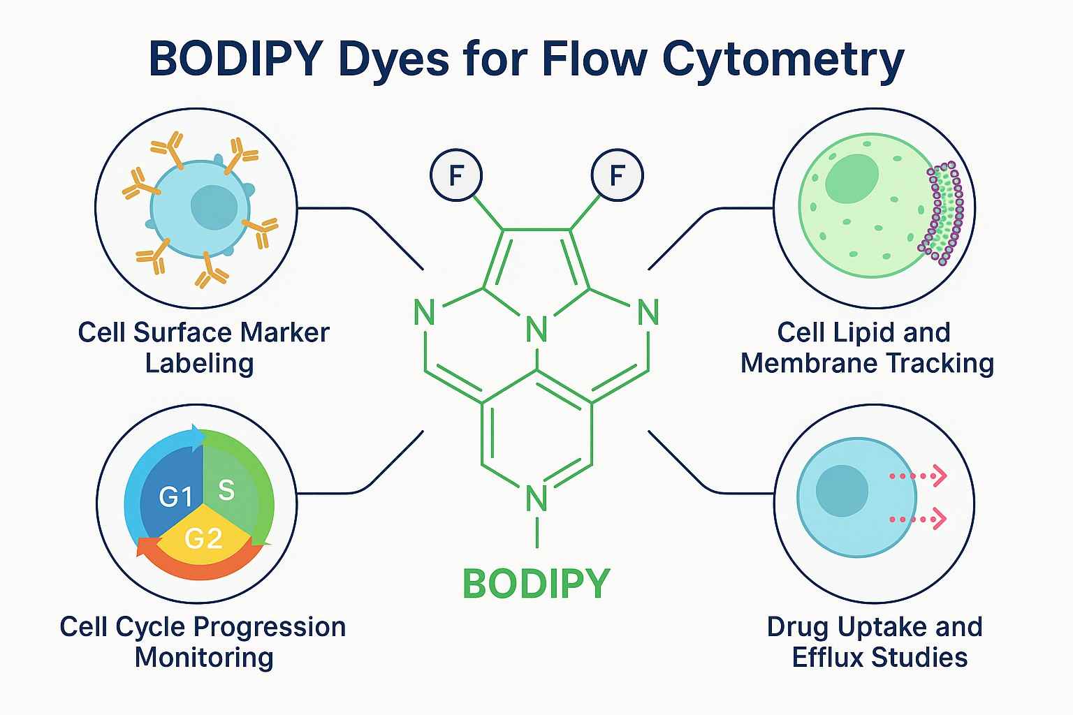
Fig. 2. BODIPY dyes for flow cytometry (BOC Sciences Authorized).
Solving Common Flow Cytometry Problems with BODIPY
As multiparameter analysis in flow cytometry continues to expand, traditional fluorescent dyes face challenges such as spectral overlap, signal bleaching, and low detection sensitivity. Thanks to their superior optical properties and structural tunability, BODIPY dyes have become a powerful solution to these bottlenecks. Their tunable emission, high photostability, high signal-to-noise ratio, and exceptional labeling ability for lipids and small molecules provide reliable support for high-resolution flow cytometry, especially for the precise detection of complex or rare cell populations.
Reduce Spectral Overlap with Tunable BODIPY Emission
The molecular structure of BODIPY dyes is highly tunable. Researchers can introduce substituents with different electronic properties to flexibly adjust the emission wavelength between 500 and 700 nm. This capability significantly reduces spectral overlap between channels, optimizes fluorescence channel distribution, minimizes compensation needs, and enhances resolution and data quality in multiparameter detection—ideal for building complex multicolor panels.
Prevent Photobleaching in Long-Term Assays
Traditional dyes are prone to fluorescence degradation under prolonged or intense laser exposure. BODIPY dyes, with their stable conjugated structure, show strong resistance to photooxidation. Even under large-scale screening or continuous excitation, they maintain stable fluorescence output, significantly reducing bleaching risks and enhancing experimental reproducibility and data reliability. They are especially suited for high-throughput cell analysis and long-term imaging experiments.
Improve Signal Resolution in Multiparameter Panels
When detecting low-abundance targets (such as rare immune cells or circulating tumor cells), signal intensity is critical. BODIPY dyes, with their high quantum yield and excellent signal-to-noise ratio, can clearly distinguish positive signals from background noise. This dramatically improves sensitivity and accuracy in rare event detection, allowing researchers to obtain more reliable biological insights.
Achieve Reliable Labeling for Difficult Targets
Unlike hydrophilic dyes, BODIPY dyes are naturally lipophilic, allowing them to penetrate cell membranes and bind efficiently to hydrophobic molecules. They can label lipid droplets, phospholipids, cholesterol, and small molecule drugs, making them widely used in lipid metabolism studies, membrane structure tracking, and drug permeability analysis. They solve the issues of low binding efficiency and poor specificity often seen with conventional dyes when labeling small molecules.
BODIPY Dyes at BOC Sciences
| Cat. No. | Product Name | CAS No. | Inquiry |
|---|---|---|---|
| F01-0012 | 3-Bodipy-propanoic acid | 165599-63-3 | Inquiry |
| F01-0194 | BODIPY FL-X SE | N/A | Inquiry |
| F01-0161 | BODIPY 558/568 C12 | 158757-84-7 | Inquiry |
| F01-0045 | BODIPY 505/515 | 21658-70-8 | Inquiry |
| F01-0171 | BODIPY 630/650 C2-Maleimide | N/A | Inquiry |
| F01-0176 | BODIPY 630/650 Cadaverine | N/A | Inquiry |
| F01-0046 | Bodipy C12-Ceramide | 1246355-58-7 | Inquiry |
| R12-0001 | BODIPY 493/503 | 121207-31-6 | Inquiry |
Applications of BODIPY in Flow Cytometry
Whether for basic immune phenotyping, lipid tracking, apoptosis analysis, or drug permeability studies, BODIPY dyes show exceptional adaptability and sensitivity, making them ideal for high-performance multiparameter flow cytometry.

Labeling of Cell Surface Markers and Intracellular Targets
BODIPY dyes can be covalently linked to antibodies, peptides, or small molecule ligands to target cell surface or intracellular biomolecules. For example, in immunophenotyping assays, BODIPY-labeled antibodies can be used to detect surface markers such as CD3 (T cells) or CD19 (B cells). They can also penetrate the cell membrane to label intracellular targets like cell cycle proteins, transcription factors, and enzymes, enabling complex phenotypic identification and pathway analysis—especially suitable for multicolor labeling experiments.

Lipid and Membrane Tracking in Cell Populations
BODIPY dyes have excellent lipophilicity and membrane affinity, often used to label membranes, lipid droplets, and phospholipid bilayers. In lipid metabolism research, they can track lipid droplet formation and distribution in real time and analyze lipid changes across different cellular states. In nanodrug carrier development, BODIPY dyes can label liposomes to monitor their release and intracellular distribution. Additionally, their fluorescence properties support high-resolution imaging, helping researchers understand dynamic behaviors such as membrane fluidity, fusion processes, and membrane protein relocation.

Monitoring Apoptosis and Cell Cycle Progression
BODIPY dyes can be combined with various biological reagents to analyze cell death and cell cycle progression. In apoptosis detection, BODIPY can be conjugated to Annexin V to detect early apoptotic signals from phosphatidylserine externalization, and used with iodide dyes (e.g., PI) to assess late apoptosis or necrosis. For cell cycle analysis, BODIPY can intercalate into DNA and be quantified by flow cytometry to distinguish cells in G1, S, and G2/M phases. This method offers high sensitivity and reproducibility, commonly used for studying anticancer mechanisms and cell proliferation activity.

Drug Uptake and Efflux Studies Using Fluorescent Probes
BODIPY dyes are frequently used to label small molecule drugs or probes, making them ideal tools for drug tracing. Their strong fluorescence and low background make them well-suited for monitoring drug uptake rate, intracellular distribution, and efflux activity. In drug resistance research, BODIPY-labeled drugs can evaluate the function of multidrug resistance pumps (e.g., P-gp); in pharmacodynamics studies, they help assess target binding efficiency and localization. This strategy not only improves visualization but also provides valuable data for targeted therapy development.
Custom BODIPY Dye Services for Flow Cytometry
BOC Sciences specializes in fluorescent dye technology and offers a wide range of customized BODIPY dyes through advanced synthesis and molecular design platforms. These services help researchers obtain clearer, more stable, and more reliable fluorescence signals in cell analysis, target detection, and mechanism studies. Key service offerings include:
Synthesis of Reactive BODIPY Derivatives
- Provides various functionalized BODIPY dyes, including NHS esters, maleimides, azides, alkynes, etc.
- Employs structurally controlled synthesis strategies to fine-tune BODIPY emission wavelengths and reactivity.
- Enables stable conjugation with common biomolecular functional groups such as amines and thiols.
- Suitable for antibody labeling, protein probe modification, and click chemistry applications.
Conjugation to Antibodies, Peptides, and Small Molecules
- Offers custom conjugation services of BODIPY dyes with antibodies, peptides, enzymes, and small molecules.
- Optimizes conjugation sites and linker lengths to preserve biomolecular functionality.
- Supports covalent labeling methods for improved dye stability and signal-to-noise ratio.
- Applied in cell surface marker detection, intracellular target localization, and targeted delivery tracking.
Hydrophilic BODIPY Variants for Aqueous Buffer Compatibility
- Incorporates hydrophilic modifications like sulfonic acid groups and PEG chains to enhance solubility and stability in aqueous systems.
- Improves compatibility with common buffers such as PBS, Tris, and HEPES.
- Reduces background noise and enhances consistency and reproducibility in flow cytometry data.
- Ideal for cell incubation assays, protein labeling, and long-term fluorescence analysis.
Multi-Color Panel Design and Dye Selection Support
- Recommends BODIPY dye combinations with non-overlapping emission wavelengths based on instrument laser and filter configurations.
- Offers channel distribution optimization strategies to reduce compensation and enhance multiparameter resolution.
- Assists customers in designing custom multicolor flow panels (3-color, 5-color, or even 8+ colors).
- Provides dye compatibility analysis, fluorescence compensation strategies, and user documentation.
Do You Need A Consultation?
BOC Sciences integrates cutting-edge fluorescence technologies to accelerate your research, driving next-generation solutions for drug discovery and diagnostics.
Versatile Fluorophores for Modern Labs
| Cat. No. | Product Name | CAS No. | Inquiry |
|---|---|---|---|
| F01-0221 | BODIPY Green 8-P2M | 929679-22-1 | Inquiry |
| F01-0220 | BODIPY TR-X NHS Ester | 217190-13-1 | Inquiry |
| F01-0215 | BODIPY 500/510, bis(SE) | N/A | Inquiry |
| F01-0163 | BODIPY FL Thapsigargin | 216571-99-2 | Inquiry |
| F01-0155 | BODIPY Fl C5-Ceramide | 133867-53-5 | Inquiry |
| F01-0181 | BODIPY 530/550 C3 | N/A | Inquiry |
| F01-0027 | BODIPY FL-X | 217190-07-3 | Inquiry |
| F01-0208 | BODIPY FL Alkyne | N/A | Inquiry |
| F01-0259 | BODIPY 16 | 2654002-78-3 | Inquiry |
| F01-0251 | BODIPY 576/589 | 150173-78-7 | Inquiry |
High-Performance Fluorescent Tools for Your Research
- TAMRA Dyes Red fluorescent labeling for biomolecule tracking.
- Cyanine5.5 Far-red fluorescence for deep tissue imaging.
- Cyanine7 Near-infrared probes for optical imaging.
- sulfo-Cyanine5.5 Water-soluble far-red imaging probe.
- sulfo-Cyanine3.5 Water-soluble orange-red fluorescent labeling.
- sulfo-Cyanine5 Hydrophilic red fluorescent bioconjugation.
- Cyanine7.5 Extended NIR imaging for in vivo studies.
- sulfo-Cyanine7.5 Hydrophilic NIR probe for bioimaging.
Explore More Topics
Online Inquiry
