

Live Cell Imaging Made Easy with Custom BODIPY Fluorophores
Cell imaging technology serves as a crucial research instrument in life sciences by allowing scientists to visually track the dynamic changes in cellular structure and function to achieve significant breakthroughs in biomedical research. Live cell imaging with traditional fluorescent dyes encounters constraints like limited photostability together with signal overlap and toxic effects, which reduce imaging quality and limit experimental adaptability. Live cell imaging now often uses BODIPY dyes because they offer superior photostability with narrow spectral emission and low toxicity levels. Custom-designed BODIPY probes enable researchers to precisely mark cell membranes and organelles as well as lipid droplets while also facilitating multimodal imaging to enhance visualization of complex biological processes.
Introduction to Cell Imaging and Its Importance in Life Sciences
Cell imaging technology, through microscopy and advanced labeling methods, helps scientists intuitively observe the complex structures inside cells and their activity processes. Particularly in the field of live cell imaging, researchers can monitor cellular physiological states, signal transduction, and metabolic dynamics in real time, thereby revealing the molecular mechanisms of disease development. Cell imaging not only promotes the development of basic biology but also provides important support for drug development, disease diagnosis, and treatment strategies. With continuous innovation in imaging technology and fluorescent probes, cell imaging is becoming more efficient and precise, greatly enriching our understanding of life phenomena.
What is Cell Imaging?
Cell imaging is a powerful technique that allows scientists to observe the structure and function of cells at the microscopic level. By using various imaging techniques, researchers can monitor biological processes inside cells in real time, such as cell division, signal transduction, metabolic activity, and cell-to-cell interactions. Cell imaging can provide not only static images of cells but also capture dynamic changes, helping us deeply understand the fundamental mechanisms of life activities. Cell imaging encompasses a variety of methods from fixed to live cells, with live cell imaging being particularly critical because it enables real-time monitoring of dynamic processes like signal transduction, protein interactions, cell migration, and metabolic changes, greatly advancing cell biology, drug development, and disease mechanism research.
Common Applications in Biological and Medical Research
Cell imaging has wide applications in biological and medical fields, including but not limited to:
- Organelle localization and functional studies: labeling mitochondria, lysosomes, Golgi apparatus, etc., to analyze their roles in cellular metabolism and signal transduction.
- Cell cycle and division monitoring: real-time observation of cell division processes and abnormalities, aiding tumor research.
- Drug screening and mechanism research: evaluating drug effects on cells and detecting drug-induced apoptosis or autophagy.
- Pathological mechanism exploration: studying viral infection, bacterial invasion, and immune cell functions, supporting disease diagnosis and treatment optimization.
- Dynamic tracking of cell signal transduction: using fluorescent probes to detect changes in signaling molecules such as calcium ions and reactive oxygen species.
The Role of Fluorescent Dyes in Visualizing Cellular Events
Fluorescent dyes are key reagents in cell imaging. By labeling specific cellular components or biomolecules, fluorescent dyes emit light of specific wavelengths, making cellular structures and dynamic processes clearly visible under a microscope. Traditional fluorescent dyes such as FITC and Rhodamine are widely used but have limitations like rapid photobleaching and weak fluorescence signals. With technological progress, new fluorescent dyes continuously emerge, bringing more possibilities to cell imaging.
Challenges in Cell Imaging Workflows
Cell imaging is essential for life science research, but practical applications face several key challenges. Traditional fluorescent dyes often suffer from poor photostability and low quantum yields, causing rapid photobleaching and weak signals, which limits long-term live cell imaging. Non-specific binding of these dyes can generate high background noise, while cytotoxic effects may disrupt normal cell functions, especially in live cells. Additionally, multiplex imaging is complicated by overlapping emission spectra of dyes, making it difficult to clearly distinguish multiple targets simultaneously. Another issue is the inconsistent permeability of dyes between live and fixed cells; some dyes easily penetrate fixed cells but fail to enter live cells without modification, adding complexity to experimental design. These technical limitations highlight the need for improved fluorescent probes with enhanced stability, specificity, low toxicity, and compatible spectral properties to advance accurate and efficient cell imaging workflows.
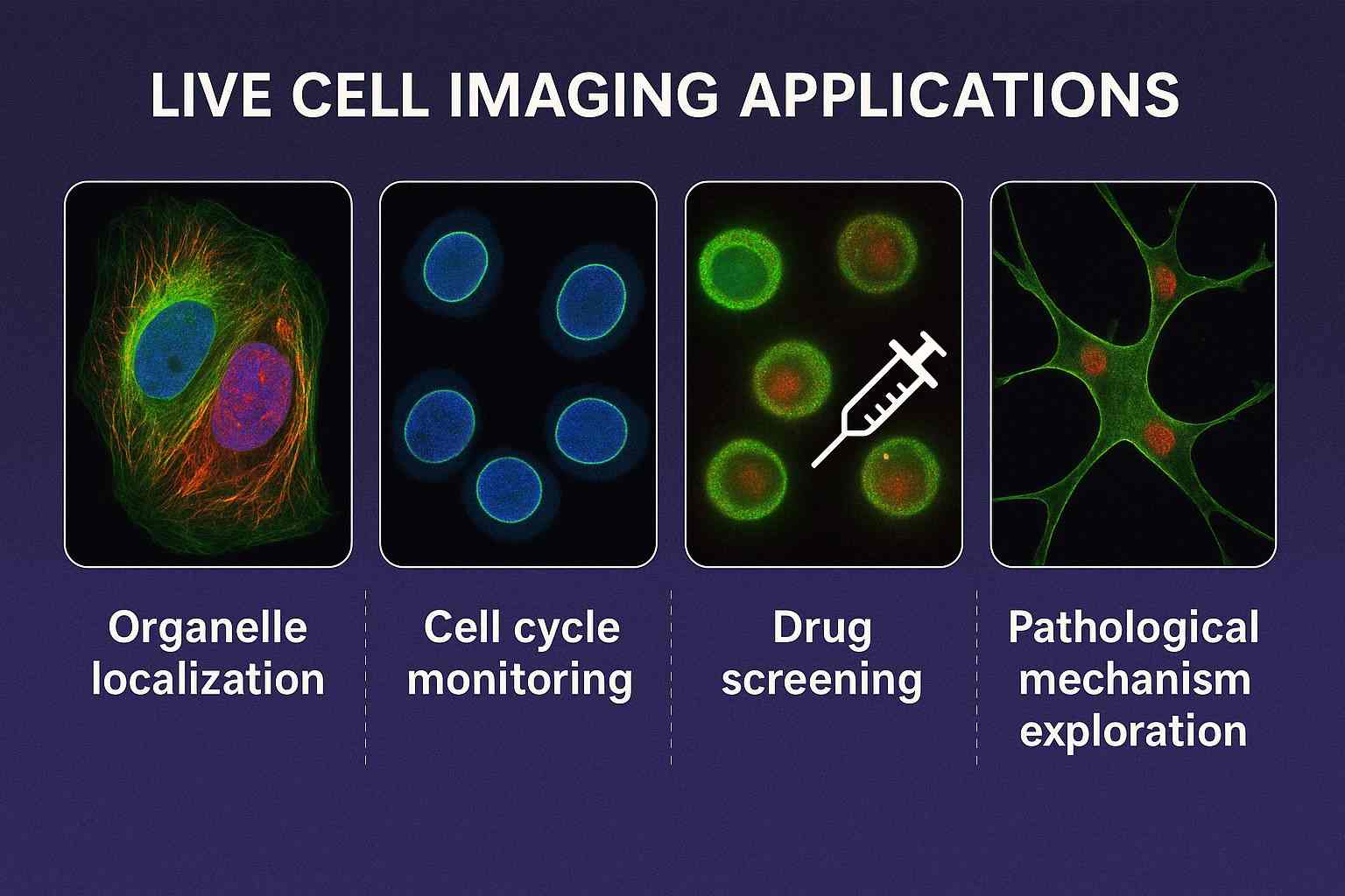
Fig. 1. Applications of live cell imaging (BOC Sciences Authorized).
Why Choose BODIPY Dyes for Cell Imaging?
BODIPY (boron-dipyrromethene) dyes, due to their unique chemical structure, exhibit excellent optical properties and biocompatibility, making them highly favored fluorescent probes in the cell imaging field in recent years. They not only have outstanding photostability and tunable emission wavelengths but also achieve low cytotoxicity and high signal-to-noise imaging effects. Moreover, BODIPY dyes are flexibly modifiable, suitable for specific labeling of various biomolecules, providing strong support for diversified cell imaging applications.
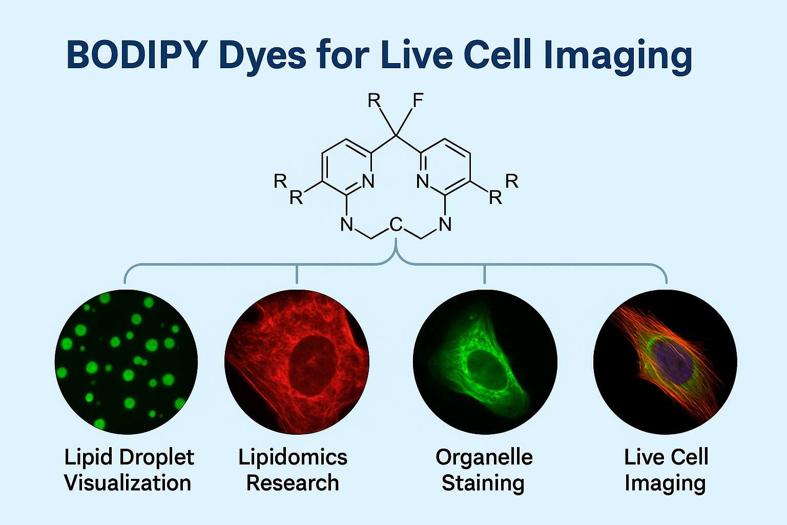 Fig. 2. BODIPY dyes for live cell imaging (BOC Sciences Authorized).
- Exceptional Photostability and Narrow Emission Peaks: BODIPY dyes are renowned for their excellent photostability, able to withstand prolonged excitation light without significant photobleaching, ensuring signal persistence and consistency during imaging. At the same time, BODIPY dyes have narrow and clear emission peaks, facilitating signal resolution in multicolor imaging and reducing spectral overlap issues.
- Tunable Fluorescence from Visible to Near-IR Regions: The chemical structure of BODIPY dyes allows molecular modifications to adjust their absorption and emission wavelengths, covering regions from green, yellow, red to near-infrared (NIR). This feature makes them suitable for different imaging equipment and experimental needs, especially in NIR imaging, which reduces background autofluorescence of biological samples and enhances imaging sensitivity.
- Low Cytotoxicity and High Signal-to-Noise Ratio: Compared to traditional dyes, BODIPY dyes have less impact on cellular bioactivity and lower cytotoxicity, making them suitable for live cell imaging. In addition, BODIPY dyes emit strong fluorescence signals with low background noise, achieving a higher signal-to-noise ratio, helping obtain clear and high-quality imaging results.
- Versatile for Labeling Lipids, Proteins, Organelles, and Nucleic Acids: BODIPY dyes can specifically bind to various biomacromolecules through chemical modification, widely used in lipid staining, protein labeling, organelle staining, and nucleic acid detection, meeting diverse cell imaging needs and helping researchers deeply analyze cellular biological processes.
BODIPY Dyes at BOC Sciences
| Cat. No. | Product Name | CAS No. | Inquiry |
|---|---|---|---|
| F01-0166 | BODIPY 493/503 NHS Ester | 216961-98-7 | Inquiry |
| F01-0215 | BODIPY 500/510, bis(SE) | N/A | Inquiry |
| F01-0152 | BODIPY-X-Alkyne | 1173281-82-7 | Inquiry |
| F01-0161 | BODIPY 558/568 C12 | 158757-84-7 | Inquiry |
| F01-0171 | BODIPY 630/650 C2-Maleimide | N/A | Inquiry |
| F01-0175 | BODIPY FL Ethylamine | N/A | Inquiry |
| F01-0046 | Bodipy C12-Ceramide | 1246355-58-7 | Inquiry |
| F01-0121 | 3-styryl-BODIPYs | 1321616-68-5 | Inquiry |
Cell Imaging Applications of BODIPY Dyes
From live cell imaging to organelle-specific staining, and to visualization of lipids and nucleic acids, BODIPY dyes have broad and diverse applications. Their excellent photostability, tunable fluorescence wavelengths, and low cytotoxicity enable them to meet various experimental needs, providing clear and accurate imaging solutions for cell biology, molecular biology, and medical research.

Live Cell Imaging with Membrane-Permeable BODIPY Probes
For live cell imaging, membrane permeability is a key parameter in fluorescent probe design. Membrane-permeable BODIPY probes specially designed for live cells can efficiently penetrate cell membranes, enter the cell interior, and selectively bind to specific targets, enabling real-time monitoring of intracellular dynamic processes. For example, BODIPY lipid probes, due to their excellent lipophilicity, can label lipid components on the cell membrane and accurately track the distribution and movement of lipids. This is significant for studying membrane protein localization and function, as well as cell signaling pathways. Through such dynamic visualization methods, researchers can gain deeper insights into cell membrane structural changes and functional regulation mechanisms.

Organelle Staining: Mitochondria, Lysosomes, and Golgi Apparatus
Functional abnormalities of organelles often underlie various diseases. By using customized BODIPY dyes for highly specific organelle staining, scientists can precisely observe morphological and functional changes of these cellular structures in live or fixed cells.
- Mitochondria Staining: BODIPY dyes can specifically enter mitochondria, helping researchers to monitor mitochondrial morphology, membrane potential, and energy metabolism status in real time. This is crucial for revealing mitochondria's role in cellular respiration and apoptosis processes.
- Lysosome Staining: Lysosomes act as the cell's "degradation factory" and play important roles in autophagy and waste processing. BODIPY dye labeling of lysosomes aids in dynamically tracking their activity and assessing the metabolic health status of cells.
- Golgi Apparatus Staining: The Golgi apparatus is responsible for protein processing, modification, and secretion. Through specific labeling by BODIPY dyes, researchers can visually observe protein transport and distribution in the Golgi apparatus, further elucidating cellular secretion mechanisms.

Lipid Droplet Visualization and Lipidomics Research
Lipid droplets are important intracellular lipid storage and metabolism sites. Their abnormal accumulation is closely associated with obesity, diabetes, fatty liver, and certain cancers. Due to their excellent lipophilicity and high fluorescence brightness, BODIPY dyes are widely used for imaging lipid droplets. Using BODIPY lipid droplet probes, researchers can precisely observe the quantity, size, and dynamic changes of lipid droplets to deeply investigate lipid metabolism and its regulatory mechanisms. Furthermore, combined with lipidomics analysis, BODIPY dyes assist in elucidating metabolic networks related to lipid droplets, providing new ideas for diagnosis and treatment of metabolic diseases.

Dual-Mode Imaging: Fluorescence and Photoacoustic Applications
Traditional fluorescence imaging offers high sensitivity and resolution but has limited penetration depth. Specially designed BODIPY dyes not only possess strong fluorescence signals but also are compatible with photoacoustic imaging technology, achieving a combination of optical and acoustic imaging. Photoacoustic imaging uses laser excitation to generate ultrasound signals, enabling deep, high-resolution imaging of tissues. When BODIPY dyes have both imaging mode capabilities, researchers can obtain more comprehensive and precise information about cells and tissues.
How to Select the Right BODIPY Dye for Your Cell Imaging Needs?
BODIPY dyes are favored for their excellent photostability, tunable fluorescence properties, and low cytotoxicity, but with numerous dye options available, choosing the best one according to experimental needs is not easy. Factors such as fluorescence wavelength, solubility, and target specificity can all affect imaging results. Additionally, comparisons with traditional fluorescent dyes and the need for customized probes also require careful consideration.
Choosing by Wavelength, Solubility, and Target Specificity
Selecting the appropriate BODIPY dye is a key step to achieving high-quality cell imaging. First, the excitation and emission wavelengths of the dye should be determined according to experimental design. For multicolor co-staining experiments, dyes with well-separated emission peaks and minimal spectral overlap should be prioritized to avoid signal confusion and cross-talk. Secondly, the dye's solubility must be considered. For cell membrane or lipid studies, lipophilic BODIPY dyes are more suitable as they effectively bind lipid components for precise labeling; hydrophilic dyes are better for labeling polar targets such as proteins or nucleic acids. Moreover, target specificity determines the dye's binding efficiency to the target structure and imaging accuracy. Choosing BODIPY probes with recognition ability toward specific targets can significantly enhance signal specificity and experimental success.
Comparing BODIPY Dyes with Other Common Fluorophores
In cell imaging, FITC and Rhodamine are widely used traditional fluorescent dyes but have some inherent drawbacks. In contrast, BODIPY dyes stand out with superior performance:
| Fluorophore | Photostability | Emission Peak Width | Cytotoxicity | Application Scope |
|---|---|---|---|---|
| FITC | Moderate | Broad | Relatively high | Fixed cells and some live cells |
| Rhodamine | Moderate | Broad | Moderate | Fixed cells and some live cells |
| BODIPY | High | Narrow | Low | Live cells and fixed cells |
Requesting Custom Probes Based on Experimental Conditions
Considering the differences in experimental environments, target types, and imaging equipment, a single dye often cannot meet all requirements. Custom BODIPY probes have become one of the solutions. By molecular structure design and chemical modification, the optical properties of dyes (such as excitation/emission wavelengths), solubility, and targeting functions can be adjusted to endow probes with stronger specificity and better imaging performance. Custom services also include optimization of reactive groups, facilitating covalent bonding with biomacromolecules or cellular components for precise labeling. Additionally, customized dyes can be tuned in their fluorescence properties according to the light source and filter characteristics of imaging devices to ensure optimal imaging results.
Custom BODIPY Dye Services from BOC Sciences
With advanced molecular design capabilities and rich synthesis experience, BOC Sciences can flexibly tune the optical properties of BODIPY dyes according to clients' specific needs, achieving precise matching of excitation and emission wavelengths. By introducing diverse reactive groups, we support efficient targeted labeling to meet the specific labeling requirements of proteins, nucleic acids, lipids, and other biomacromolecules.
Structure Design and Fluorophore Tuning
- Flexibly design BODIPY core structures according to customer requirements to achieve precise tuning of excitation and emission wavelengths, covering visible to near-infrared ranges.
- Optimize dye photostability and quantum yield by introducing various substituents, enhancing imaging signal strength and durability.
- Adjust dye polarity and hydrophobicity to ensure probes adapt to different biological environments and cell types, improving membrane permeability and targeting ability.
- Customize multifunctional fluorophores suitable for multiplex staining and complex imaging platforms by combining multiple photophysical property requirements.
Reactive Group Modification for Target-Specific Labeling
- Design and introduce diverse reactive groups (e.g., amino, carboxyl, azide, thiol) to facilitate covalent bonding with biomacromolecules.
- Support specific labeling of proteins, nucleic acids, lipids, and small molecule targets, meeting imaging needs for various research subjects.
- Optimize reactive group stability and reaction conditions to ensure efficient, mild, and cell-friendly labeling processes.
- Provide customized linker designs to achieve reasonable spatial arrangement, enhancing labeling efficiency and fluorescence signal quality.
Batch-to-Batch Consistency and Scale-Up Support
- Strictly control synthesis processes to ensure high consistency of purity, optical performance, and chemical properties between batches, guaranteeing reproducibility of experimental data.
- Possess advanced production facilities and complete quality management systems, supporting smooth transition from small-scale R&D samples to large-scale industrial production.
- Offer flexible capacity support to meet diverse needs in different R&D stages and commercialization applications.
- Equipped with comprehensive quality testing and traceability systems, ensuring each batch meets high-quality standards.
Technical Support for Probe Integration in Imaging Platforms
- Provide professional technical guidance to assist clients in successfully integrating custom BODIPY probes into various cell imaging platforms and experimental workflows.
- Optimize dye usage concentration, staining time, and imaging parameters according to client experimental conditions to enhance imaging effects and data quality.
- Offer probe labeling scheme design based on client needs, including co-staining, multichannel imaging, and live cell dynamic observation technical support.
- Continuously follow up technical services to solve client application problems, ensuring smooth project progress.
Do You Need A Consultation?
BOC Sciences integrates cutting-edge fluorescence technologies to accelerate your research, driving next-generation solutions for drug discovery and diagnostics.
Versatile Fluorophores for Modern Labs
| Cat. No. | Product Name | CAS No. | Inquiry |
|---|---|---|---|
| F01-0046 | Bodipy C12-Ceramide | 1246355-58-7 | Inquiry |
| F01-0180 | BODIPY 630/650 Acid | N/A | Inquiry |
| F01-0182 | BODIPY 558/568 C3 | N/A | Inquiry |
| F01-0121 | 3-styryl-BODIPYs | 1321616-68-5 | Inquiry |
| F01-0044 | BODIPY-Cholesterol | 878557-19-8 | Inquiry |
| F01-0029 | BODIPY 558/568; bodipy-SE | N/A | Inquiry |
| F01-0037 | azido-Bodipy-650 | N/A | Inquiry |
| F01-0257 | C11 BODIPY 581/591 | 217075-36-0 | Inquiry |
| F01-0251 | BODIPY 576/589 | 150173-78-7 | Inquiry |
| F01-0216 | BODIPY 576/589, Acid | N/A | Inquiry |
High-Performance Fluorescent Tools for Your Research
- JOE Dyes Green fluorescent probes for qPCR applications.
- Cyanine7 Near-infrared probes for optical imaging.
- ATTO Dyes High-performance fluorophores for super-resolution microscopy.
- sulfo-Cyanine7 Water-soluble near-infrared fluorescent labeling.
- Cyanine3 Standard green-orange fluorescent biomolecular labeling.
- Cyanine7.5 Extended NIR imaging for in vivo studies.
- Coumarin Blue fluorescent probes for enzyme assays.
- BODIPY Photostable dyes for lipid and cell imaging.
Explore More Topics
Online Inquiry
