

Flow Cytometry – Intracellular and Nuclear Factor Staining Protocol
Staining and detecting intracellular molecules helps identify cell subpopulations and states. Unlike traditional surface antibody staining, flow cytometry analysis of intracellular proteins requires initial fixation to stabilize the cell membrane, followed by permeabilization to allow antibodies to access intracellular antigens. Mass cytometry extends this capability by simultaneously analyzing both surface markers and intracellular or nuclear components across multiple channels, without the need for voltage adjustment or compensation, greatly enhancing detection power.
Intracellular Factor Staining Protocol
Intracellular Cytokine Staining (ICCS) is a key technique for evaluating immune cell functional activity and differentiation states, such as identifying Th1, Th2, and Th17 subpopulations. Unlike constitutively expressed structural proteins, most cytokines are synthesized only after cell activation and are rapidly secreted, resulting in very low intracellular levels that are difficult to detect. Therefore, the core strategy of this protocol involves induction and blockade: cells are first stimulated in vitro (e.g., with PMA/ionomycin) to initiate cytokine production while protein transport inhibitors (e.g., Brefeldin A) block secretion, causing the cytokines to accumulate in the cytoplasm. Subsequent fixation and permeabilization allow precise quantitative analysis of specific functional proteins at the single-cell level.
Step-by-Step ICCS Workflow:
- Cell Activation and Stimulation: Resting T and B cells express very low levels of cytokines and require in vitro activation. Common stimulants include PMA and ionomycin, which jointly activate PKC and induce T cell activation. Protein transport inhibitors (e.g., Brefeldin A or monensin) are added during stimulation to prevent cytokine secretion and retain intracellular accumulation. Typically, cells are stimulated for 4–6 hours at 37°C with 5% CO₂.
- Cell Fixation: For combined surface marker staining, perform surface staining first. Then add fixation buffer and incubate at room temperature in the dark for 20 minutes. After centrifugation and supernatant removal, cells can be temporarily stored at 4°C or −80°C in an appropriate preservation solution.
- Cell Permeabilization: Resuspend fixed cells in 1× permeabilization wash buffer, centrifuge at 350×g for 5–10 minutes, discard supernatant, and wash once more. Permeabilization allows antibodies to enter the cell and bind target antigens.
- Intracellular Staining: Resuspend cells in 1× permeabilization wash buffer, add the corresponding antibody, and incubate at room temperature in the dark for 20 minutes. If using biotinylated antibodies, follow with a streptavidin–fluorophore secondary antibody incubation. Wash twice and discard the supernatant.
- Sample Preparation and Analysis: Finally, resuspend cells in cell staining buffer, set up controls, and perform flow cytometry. To ensure specificity, blocking experiments with unlabeled antibodies or recombinant cytokines can be performed to rule out non-specific binding.
Nuclear Factor Staining Protocol
Nuclear transcription factors (e.g., FoxP3, Ki-67, T-bet) are key proteins that determine cell lineage differentiation and functional state. Unlike cytoplasmic cytokine staining, nuclear factor detection requires antibodies to penetrate both the cell and nuclear membranes, necessitating stricter fixation and permeabilization conditions. This protocol uses a specialized transcription factor fixation/permeabilization buffer system designed to allow antibodies to access nuclear antigens while preserving chromosomal DNA integrity and nuclear protein epitopes, ensuring high signal-to-noise flow cytometry results.
Step-by-Step Nuclear Factor Staining Workflow:
- Aliquot 100 μL of cells per tube (~1×10⁶ cells).
- (Optional) Stain with fixable viability dye according to instructions.
- (Optional) Perform Fc receptor blocking if required.
- Stain surface markers with fluorescently labeled antibodies according to the antibody instructions.
- After incubation, add 1 mL cell staining buffer, centrifuge at 300×g for 5 min, discard supernatant, and resuspend cells in 100 μL cell staining buffer.
- Add 1 mL 1× fixation working solution, vortex to mix, incubate at 4°C for 30 min, then centrifuge at 600×g for 5 min and discard supernatant.
- Add 2 mL 1× permeabilization working solution, vortex, centrifuge at 600×g for 5 min, discard supernatant.
- Repeat step 7.
- Resuspend cells in 100 μL 1× permeabilization working solution.
- Add fluorescently labeled nuclear antibodies per the instructions, mix, and incubate at room temperature in the dark for 30 min.
- Add 2 mL 1× permeabilization working solution, centrifuge at 600×g for 5 min, discard supernatant.
- Resuspend cells in an appropriate volume of cell staining buffer for flow cytometry analysis.
Case Study: Accurate Identification of Regulatory T Cells (Tregs, CD4⁺ CD25⁺ FoxP3⁺)
Regulatory T cells (Tregs) are essential for maintaining immune tolerance. While CD4 and CD25 are common surface markers, activated effector T cells can also upregulate CD25 during inflammation, making surface staining alone insufficient to distinguish true Tregs.
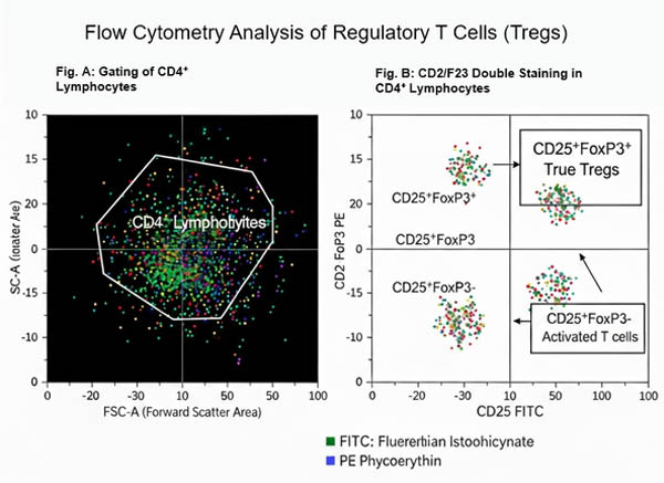 Fig. 1. Flow cytometry analysis of regulatory T cells (Tregs) (BOC Sciences Authorized).
Detection Strategy and Key Details:
- Multidimensional Markers: This approach combines surface markers (CD4, CD25) with nuclear transcription factor (FoxP3) staining. FoxP3 is the master regulator of Treg development and function and is currently the most specific Treg identifier.
- Critical Steps: Surface staining for CD4 and CD25 is performed first, followed by fixation and permeabilization using a dedicated nuclear factor kit, and then FoxP3 antibody incubation.
- Data Analysis: In flow cytometry scatter plots, gating on CD4⁺ cells reveals a clear CD25⁺FoxP3⁺ population. Compared with surface staining alone, including FoxP3 eliminates false positives (e.g., CD25⁺FoxP3⁻ activated T cells) and allows functional assessment of Tregs via FoxP3 mean fluorescence intensity (MFI). This strategy is widely applied in autoimmune disease studies, tumor microenvironment analysis, and transplantation immunology research.
Fluorescent Dyes Recommended for Your Research Project
| Catalog | Name | CAS | Inquiry |
|---|---|---|---|
| F04-0012 | FITC isomer I | 3326-32-7 | Bulk Inquiry |
| A16-0004 | Phalloidin-FITC | 915026-99-2 | Bulk Inquiry |
| F02-0030 | Cy3-NHS ester | 146368-16-3 | Bulk Inquiry |
| A16-0074 | Biotin DHPE | 136235-58-0 | Bulk Inquiry |
| A16-0092 | Biotin-XX-SSE | 194041-66-2 | Bulk Inquiry |
| R15-0017 | TCO-PEG4-biotin | 2183440-30-2 | Bulk Inquiry |
| F07-0010 | Biotin-PEG4-Dde-TAMRA-PEG3-Azide | 2353409-56-8 | Bulk Inquiry |
| F04-0032 | FAM isothiocyanate (FITC), 5- and 6-isomers | 27072-45-3 | Bulk Inquiry |
| F07-0016 | Dde TAMRA Biotin Alkyne | 2353409-55-7 | Bulk Inquiry |
| F03-0024 | diSulfo-Cy3 azide | 2055138-89-9 | Bulk Inquiry |
| A19-0101 | Biotin-Aniline | 769933-15-5 | Bulk Inquiry |
| A19-0104 | TO1-Biotin | 1622455-90-6 | Bulk Inquiry |
| R01-0042 | AF594 activated ester, 5-isomer | 1638544-48-5 | Bulk Inquiry |
| R01-0471 | AF647 NHS ester | 407627-60-5 | Bulk Inquiry |
| R01-0451 | AF 488 TFP ester | 2133404-55-2 | Bulk Inquiry |
| A17-0130 | N-methyl-N'-(azide-PEG3)-Cy3 | 2107273-16-3 | Bulk Inquiry |
| F03-0023 | Sulfo-Cy3-Methyltetrazine | 1801924-47-9 | Bulk Inquiry |
| F03-0025 | Trisulfo-Cy3-Alkyne | 1895849-34-9 | Bulk Inquiry |
Corporate Profile
BOC Sciences provides global research and industry clients with a wide range of fluorescent probes, dyes, and custom solutions. Built on a strong platform in chemical synthesis and functional materials, we offer a complete portfolio covering small-molecule fluorescent dyes, labeling reagents, in vivo imaging probes, and cellular and molecular imaging tools. Our products are widely used in life science research, bioimaging, diagnostic development, and advanced materials science.
Through probes by BOC Sciences, our specialized subsite, we focus on high-performance fluorescent probes and innovative labeling technologies. You can find conventional dyes like FITC, TRITC, Cy series, Alexa Fluor series, near-infrared probes, environment-responsive probes, and targeted fluorescent molecules. We also provide custom services such as structural modification, spectral tuning, conjugation, and tailored synthesis to meet your research needs.
Our team of experienced chemists and biologists works with advanced synthesis, purification, and analytical equipment, ensuring every product delivers consistent purity, stability, and optical performance. BOC Sciences' probes and services are trusted by universities, research institutes, and biopharmaceutical companies worldwide for cellular imaging, molecular tracing, mechanism studies, and method development. We aim to be your long-term partner in fluorescent labeling and imaging research.
Custom Fluorescent Solutions Designed for Your Experiments
- DNA StainingPrecise fluorescent dyes for clear DNA visualization and analysis.
- Lipid StainingFluorescent solutions for effective lipid structure imaging.
- Cell StainingAdvanced staining for detailed cell morphology and analysis.
- Protein StainingHigh-quality staining for accurate protein detection and imaging.
- Bacteria ImagingFluorescent solutions to visualize bacterial structures and activity.
- Cell ImagingVisualize and analyze live or fixed cells using advanced fluorescence.
- Molecular ImagingCutting-edge fluorescent solutions for deep molecular analysis.
- Fluorescence ImagingHigh-resolution imaging solutions for detailed fluorescence studies.
- BioconjugationCustom bioconjugation services for protein, peptide, and dye linking.
- Drug DeliveryTailored fluorescent solutions for efficient drug delivery research.
- Molecular DiagnosticsFluorescent probes and markers for precise molecular diagnostics.
- Flow CytometryFluorescent dyes and reagents for enhanced flow cytometry analysis.
High-Performance Fluorescent Tools for Your Research
- Apoptosis Fluorescent Probes Probes detecting programmed cell death events.
- Nuclear Fluorescent Probes DNA-binding dyes for nucleus visualization.
- Golgi Fluorescent Probes Targeted probes for Golgi apparatus visualization.
- Cell Proliferation Tracer Fluorescent Probes Long-term tracking of cell division processes.
- Lipid Fluorescent Probes Dyes for lipid droplets and membranes.
- pH Indicators Fluorescent sensors for intracellular pH monitoring.
- Metal Fluorescent Probes Selective sensors for intracellular metal ions.
- Lysosomal Fluorescent Probes Acidic organelle markers for lysosome tracking.
Explore More Topics
Online Inquiry
