

Lipid Staining – Guide to Lipid Droplet & Cholesterol Staining Protocols
Lipids are a class of organic molecules widely present in living organisms, playing essential roles in cellular structure and function. Lipid droplets and cholesterol represent different forms of lipids with distinct structural and functional characteristics. Lipid droplets serve as the primary storage sites for neutral lipids, including triglycerides and cholesteryl esters. Under pathological conditions, such as organ steatosis, increased intracellular lipid droplets may appear, which can be clearly visualized by lipid staining. Cholesterol is an essential component of eukaryotic cell membranes, maintaining membrane integrity, regulating membrane fluidity, and participating in signal transduction. It plays critical roles in the development and progression of diseases such as atherosclerosis, Niemann-Pick disease type C (NPC), and Alzheimer's disease.
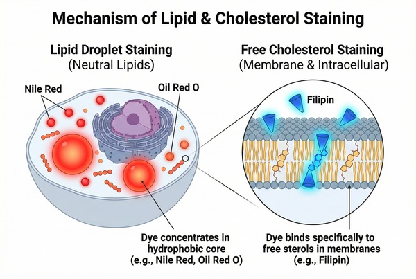 Fig. 1. Cellular mechanisms of lipid droplet and free cholesterol staining (BOC Sciences Authorized).
Staining of lipid droplets and cholesterol is an important technique in pathology and cell biology research for observing and quantifying intracellular lipid accumulation. Currently, the most widely used dyes for lipid droplet staining include Oil Red O, Nile Red, and Sudan dyes. For cholesterol staining, the most commonly used dye is Filipin III, which has been widely applied for more than 30 years. It can label free cholesterol in biological membrane structures and non-cellular structures. In combination with cholesterol esterase, cholesteryl esters can be hydrolyzed into free cholesterol for indirect visualization.
Comparison Table of Different Dye Selection
The table below helps researchers quickly select appropriate dyes based on experimental requirements (e.g., live cells vs. fixed cells).
| Dye Name | Target Lipid | Detection Method | Advantages | Limitations |
|---|---|---|---|---|
| Oil Red O | Neutral lipids / triglycerides | Bright-field microscopy | Classic method, low cost, visually intuitive | Requires frozen sections; not suitable for live-cell tracing |
| Nile Red | Lipid droplets / phospholipids | Fluorescence / Flow cytometry | Extremely high sensitivity; multiple color options available | Broad emission spectrum; prone to spectral overlap |
| Filipin III | Free cholesterol | Fluorescence (UV) | Highly specific for cholesterol | Easily quenched; UV excitation may cause cellular damage |
| BODIPY-Chol | Cholesterol dynamics | Fluorescence (Green) | Compatible with live cells; good photostability | Cholesterol analog; potential metabolic interference should be considered |
Lipid Droplet Staining Protocol
Tissue Lipid Droplet Staining with Oil Red O
Principle: Oil Red O is an azo dye with strong lipid solubility and lipid-staining capability. It binds to triglycerides, forming small lipid droplet-like deposits. Lipophilic dyes dissolve in lipids within tissues and cells, and their solubility in lipids is higher than in the solvent. When tissue sections are immersed in the staining solution, the dye leaves the solution and dissolves into lipids within the tissue (such as lipid droplets), rendering them orange-red.
Staining Procedure:
1) Preparation of Oil Red O staining solution:
a. Stock solution: Weigh 1 g Oil Red O and dissolve in 100 mL isopropanol to prepare a stock solution. Store in a brown bottle at 4°C, sealed and protected from light.
b. Working solution: Before use, dilute the stock solution to 60% with double-distilled water. Filter through filter paper before use.
2) Cut 5 μm-thick liver tissue sections using a cryostat;
3) Fix with 95% ethanol, rinse with distilled water for 1 min, and immerse in 60% isopropanol for 30 min;
4) Stain with 60% Oil Red O solution for 10–15 min, then rinse with distilled water for 1 min;
5) Counterstain with hematoxylin, rinse with tap water for 1–3 min, and observe lipid deposition in hepatocytes under a light microscope.
Oil Red O Staining Results of Liver Tissue:
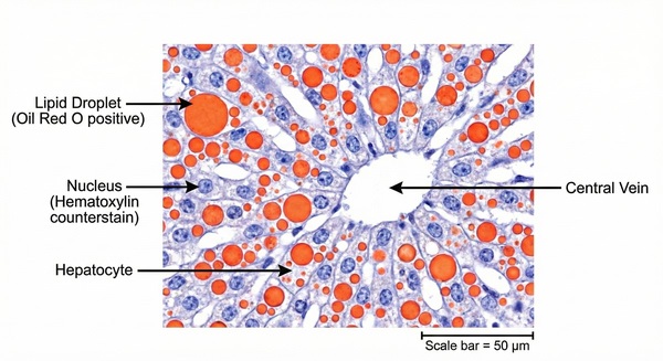 Fig. 2. Oil Red O staining of lipid droplets in hepatocytes (BOC Sciences Authorized).
Product Applications:
- Clinical pathological diagnosis: Commonly used for rapid auxiliary diagnosis of fat embolism, hepatic steatosis (such as NAFLD/NASH), renal clear cell carcinoma, and certain lipid metabolic diseases.
- Metabolic disease research: Observation of lipid accumulation in liver, skeletal muscle, or brown adipose tissue in obesity and diabetes models.
- Atherosclerosis research: Used for en face staining of the aorta or frozen sections of the aortic root to quantify lipid distribution in atherosclerotic plaques.
Cellular Lipid Droplet Staining with Nile Red
Principle: Nile Red is a lipophilic fluorescent dye and an environment-sensitive fluorophore. It exhibits strong fluorescence in lipid-rich environments while showing minimal fluorescence in aqueous media. This property makes it an important fluorescent indicator for detecting intracellular lipid droplets. Results can be analyzed using fluorescence microscopy or flow cytometry.
Staining Procedure:
1) Preparation of Nile Red staining solution:
a. Stock solution: Dissolve an appropriate amount of Nile Red (Mw: 318.37 g/mol) in anhydrous DMSO to prepare a 1 mM stock solution. Aliquot according to single-use volumes, store frozen, avoid repeated freeze-thaw cycles, and protect from light.
b. Working solution: Dilute the stock solution 1:1000 in HHBS or physiological buffer (pH 7.0) to obtain 1× Nile Red working solution. Vortex to mix well.
[Note]: The actual staining concentration should be optimized according to literature or laboratory conditions. The above is for reference only.
2) Treat cells with test compounds for an appropriate period;
3) Centrifuge and adjust cell density to 1–5 × 10^5 cells/tube;
4) Resuspend cells in 500 μL Nile Red working solution;
5) Incubate at room temperature or 37°C for 5–10 min in the dark;
6) Remove staining solution and wash cells with HHBS or appropriate buffer;
7) Resuspend cells in 500 μL pre-warmed HHBS or culture medium to achieve a density of 1–5 × 10^5 cells/tube;
8) Detect fluorescence signals by fluorescence microscopy or flow cytometry (Ex/Em = 552/636 nm).
[Notes]:
a. For adherent cells, wash with HHBS or appropriate buffer and directly add Nile Red working solution for incubation;
b. Cells may also be pre-fixed before staining with Nile Red.
Product Applications:
- Lipid metabolism regulation: Study dynamic changes in lipid droplet formation in various cells (e.g., adipocytes, hepatocytes, macrophage-derived foam cells) under induction or drug intervention.
- High-throughput screening: Combined with flow cytometry or high-content screening systems to screen large-scale compound libraries for anti-obesity or lipid secretion-regulating agents.
- Environmental toxicology monitoring: Use Nile Red staining in aquatic organisms or indicator cells to assess the effects of environmental pollutants (e.g., microplastics, endocrine disruptors) on lipid metabolism.
Cholesterol Staining Protocol
Cellular Cholesterol Staining with Filipin III
Principle: The main component of Filipin is Filipin III, which specifically interacts with free cholesterol but not with esterified cholesterol. This specificity arises from multiple conjugated double bonds in its molecular structure, enabling complex formation with the hydrophobic region of cholesterol. After binding to free cholesterol, Filipin III exhibits an excitation peak at approximately 360 nm and an emission peak at approximately 480 nm. Detection of fluorescence signals near these wavelengths allows characterization of intracellular cholesterol content.
Staining Procedure:
1) Preparation of Filipin staining solution:
a. Stock solution: Add 1 mL DMSO to 1 mg Filipin powder to prepare a 1 mg/mL stock solution. Aliquot for single use, store frozen, avoid repeated freeze-thaw cycles, and protect from light.
b. Working solution: Before use, dilute the stock solution in PBS to the desired concentration (e.g., 0.1 mg/mL).
2) Seed cells on coverslips and culture for 12–18 h;
3) Wash three times with PBS and fix with 4% paraformaldehyde at room temperature for 20 min;
4) Incubate cells with 0.1 mg/mL Filipin III at room temperature for 30 min;
5) Counterstain with 0.35 μg/mL propidium iodide (PI) for 5 min;
6) Detect fluorescence intensity of Filipin and PI using a confocal laser scanning microscope with 405 nm excitation.
Filipin Staining Results in Astrocytes/Neurons:
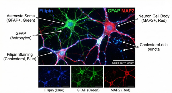 Fig. 3. Filipin staining showing free cholesterol in neuron-astrocyte co-culture (BOC Sciences Authorized).
Product Applications:
- Rare disease screening: Filipin staining is the gold standard cytological method for diagnosing Niemann-Pick disease type C (NPC), by observing abnormal accumulation of free cholesterol in lysosomes of fibroblasts.
- Cholesterol homeostasis research: Study regulation of intracellular cholesterol levels by the SREBP/SCAP signaling pathway and the distribution of cholesterol between the plasma membrane and intracellular organelles.
- Viral infection mechanism research: Investigate the mediating role of host membrane cholesterol in infections by enveloped viruses such as HBV and SARS-CoV-2.
BODIPY-Cholesterol Staining
Principle: BODIPY-Cholesterol is cholesterol labeled with the BODIPY (boron-dipyrromethene) fluorescent group. It possesses biological activity and membrane permeability, allowing cellular uptake and intracellular localization tracking of cholesterol metabolism. It is commonly used as a fluorescent probe to monitor cholesterol uptake and organelle-to-organelle cholesterol trafficking. In addition to its fluorescent properties, BODIPY-cholesterol retains the biological activity of cholesterol. It can mimic the behavior of native cholesterol and interact with intracellular cholesterol receptors, thereby participating in normal physiological processes.
Staining Procedure:
1) Preparation of BODIPY-Cholesterol staining solution:
a. Stock solution: Dissolve an appropriate amount of BODIPY-Cholesterol solid in anhydrous ethanol or DMSO to prepare a 1–5 mM stock solution. Aliquot in small volumes and store at −20°C protected from light.
b. Working solution: Before the experiment, dilute the stock solution to a final concentration (commonly 1–5 μM) in pre-warmed serum-free culture medium or HBSS buffer.
[Note]: Cholesterol probes tend to aggregate. It is recommended to add fatty acid-free BSA (final concentration 0.1%–0.5%) to improve solubility.
2) Cell preparation: Seed cells on coverslips or fluorescence-compatible culture dishes and allow them to reach 60–70% confluence;
3) Drug treatment (optional): Treat cells with test compounds as required;
4) Probe incubation: Remove old medium, wash twice with PBS, add prepared BODIPY-Cholesterol working solution, and incubate at 37°C with 5% CO₂ for 15–30 min in the dark;
5) Washing: Discard staining solution and gently wash three times with pre-warmed PBS or HBSS to remove unbound probe;
6) Fixation (optional):
a. Live-cell imaging: Directly add phenol red-free imaging buffer for real-time observation.
b. Fixed-cell imaging: Fix with 4% paraformaldehyde at room temperature for 15 min, wash with PBS, and proceed to subsequent steps.
[Note]: BODIPY probes are soluble in organic solvents. After fixation, do not use permeabilization buffers containing Triton X-100 or mounting media containing ethanol.
7) Observation and detection: Observe using a fluorescence or confocal microscope. Excitation/emission wavelengths: Ex/Em = 491/508 nm (green fluorescence, similar to FITC/GFP channel).
Product Applications:
- Basic research: Investigate abnormal cellular cholesterol uptake in models of metabolic diseases and atherosclerosis;
- Drug screening: Rapidly screen candidate drugs or inhibitors that regulate cholesterol absorption;
- Mechanistic studies: Real-time tracking of dynamic cholesterol transport between organelles.
Fluorescent Dyes Recommended for Your Research Project
| Catalog | Name | CAS | Inquiry |
|---|---|---|---|
| A16-0159 | HIDC iodide | 36536-22-8 | Bulk Inquiry |
| A16-0201 | DAPI dihydrochloride | 28718-90-3 | Bulk Inquiry |
| A16-0082 | DiOC16(3) | 161433-32-1 | Bulk Inquiry |
| A16-0084 | Speed DiO | 164472-75-7 | Bulk Inquiry |
| A16-0111 | BF 594 Phalloidin | 330626-83-0 | Bulk Inquiry |
| A16-0003 | Phalloidin-TFAX 488 | 289620-19-5 | Bulk Inquiry |
| A16-0002 | Phalloidin-TRITC | 915013-10-4 | Bulk Inquiry |
| A16-0166 | 3,3'-Diheptylthiacarbocyanine iodide | 53213-88-0 | Bulk Inquiry |
| A16-0091 | Lucifer yellow CH ammonium salt | 188904-20-3 | Bulk Inquiry |
| A16-0163 | 3,3'-Dipropyloxacarbocyanine iodide | 53213-79-9 | Bulk Inquiry |
| A16-0008 | DiD perchlorate | 127274-91-3 | Bulk Inquiry |
| A16-0212 | DiO, lipophilic tracer | 28462-56-8 | Bulk Inquiry |
Corporate Profile
As a premier partner in life science research, BOC Sciences provides comprehensive, state-of-the-art fluorescent imaging solutions that empower scientists to visualize the microscopic world with exceptional clarity. Building upon our advanced expertise in specialized lipid and cholesterol tracking, we offer a vast, high-quality portfolio of fluorescent dyes engineered for precise subcellular localization and dynamic analysis.
Our extensive product line covers every critical cellular compartment. We provide highly specific and reliable stains, including membrane probes (e.g., DiI, DiO, DiR), nuclear dyes (e.g., DAPI, Hoechst 33342), and cytoskeleton labels (e.g., FITC-Phalloidin). Additionally, we offer robust organelle-specific trackers for mitochondria (e.g., JC-1, Rhodamine 123), lysosomes (e.g., Lyso-Tracker Red/Green), the endoplasmic reticulum (e.g., ER-Tracker series), and exosomes. Whether you are conducting routine fixed-cell imaging or complex live-cell dynamic studies, our reagents ensure superior brightness, photostability, and minimal background.
Beyond our off-the-shelf catalog, our core strength lies in our robust team capabilities and end-to-end support services. Our seasoned R&D chemists and biologists specialize in custom fluorophore synthesis and targeted bioconjugation, tailoring solutions to your exact experimental needs. Furthermore, our dedicated technical experts provide hands-on application support—helping you optimize staining protocols, troubleshoot complex imaging assays, and accelerate your discoveries in disease modeling and high-throughput drug screening. At BOC Sciences, we are committed to illuminating your research through innovative products and unwavering scientific partnership.
Custom Fluorescent Solutions Designed for Your Experiments
- DNA StainingPrecise fluorescent dyes for clear DNA visualization and analysis.
- Lipid StainingFluorescent solutions for effective lipid structure imaging.
- Cell StainingAdvanced staining for detailed cell morphology and analysis.
- Protein StainingHigh-quality staining for accurate protein detection and imaging.
- Bacteria ImagingFluorescent solutions to visualize bacterial structures and activity.
- Cell ImagingVisualize and analyze live or fixed cells using advanced fluorescence.
- Molecular ImagingCutting-edge fluorescent solutions for deep molecular analysis.
- Fluorescence ImagingHigh-resolution imaging solutions for detailed fluorescence studies.
- BioconjugationCustom bioconjugation services for protein, peptide, and dye linking.
- Drug DeliveryTailored fluorescent solutions for efficient drug delivery research.
- Molecular DiagnosticsFluorescent probes and markers for precise molecular diagnostics.
- Flow CytometryFluorescent dyes and reagents for enhanced flow cytometry analysis.
High-Performance Fluorescent Tools for Your Research
- pH Indicators Fluorescent sensors for intracellular pH monitoring.
- Mitochondrial Fluorescent Probes Targeted dyes for mitochondrial structure and function.
- Nerve Terminal Probes Fluorescent tracers for synaptic activity analysis.
- Calcium, Chloride and Other indicators Fluorescent indicators for intracellular ion flux monitoring.
- Cell Proliferation Tracer Fluorescent Probes Long-term tracking of cell division processes.
- Nuclear Fluorescent Probes DNA-binding dyes for nucleus visualization.
- Ion Fluorescent Probes Indicators for real-time ion concentration imaging.
- Nitric Oxide (NO) & Reactive Oxygen Species (ROS) Probes for oxidative stress and signaling detection.
Explore More Topics
Online Inquiry
