

How to Choose a Fluorophore for Fluorescent Labeling Experiments?
Choosing a fluorophore is one of the most consequential decisions in a fluorescent labeling workflow. In many experiments, the fluorescent signal itself is not the main problem. The real challenge is selecting a label that produces usable signal under the right excitation source, fits the detection platform, remains compatible with the target molecule, and does not create avoidable interpretation problems in downstream analysis. A fluorophore that looks attractive in a catalog can perform poorly in practice if its spectra do not match the instrument, if it bleaches too quickly, if it adds too much hydrophobicity to the target, or if it complicates multicolor design. This is why fluorophore selection should be treated as a design step rather than a final shopping decision.
The best fluorophore is therefore not simply the brightest one or the most commonly used one. It is the one that fits the full experimental context. That context includes the target being labeled, the chemistry used to attach the fluorophore, the signal duration required, the likelihood of background or spectral spillover, and the practical limits of purification and validation. In a protein-labeling project, the main issue may be preserving binding or activity after conjugation. In nucleic acid labeling, quenching, position effects, and readout design may matter more. In multicolor imaging or flow workflows, channel separation and instrument compatibility can become more important than raw brightness alone.
Why Fluorophore Choice Matters?
Fluorophore choice influences much more than the color of the final signal. It affects whether the signal can be excited efficiently, whether it remains visible across the intended observation window, whether it can be separated from other channels, and whether the target still behaves in a meaningful way after labeling. A poor fluorophore match often does not produce total failure. Instead, it produces weak, unstable, crowded, or misleading data that require additional controls and complicate interpretation. This is why fluorophore selection belongs near the beginning of experimental planning rather than being treated as an interchangeable downstream detail.
- Signal Quality Starts with Fluorophore Fit: Signal quality depends on how well the fluorophore fits the biological and technical context. A dye that performs well in a simple purified conjugate may not perform equally well in dense cell imaging, long time-lapse acquisition, or a multiplex panel where neighboring channels already compete for spectral space. Good fluorophore fit means the excitation source can drive the signal efficiently, the detector can collect it cleanly, and the label remains interpretable under real workflow conditions rather than idealized single-channel testing. In practice, this is often the difference between a fluorophore that produces an attractive pilot result and one that remains reliable through the full experiment.
- Detection Performance Depends on More Than Brightness: Brightness matters, but brightness alone does not guarantee a strong experiment. A bright fluorophore can still be a poor choice if it photobleaches quickly, aggregates on the target, introduces high background, or produces severe spectral overlap with more important channels. Conversely, a fluorophore with moderate brightness may be the better option when it has cleaner spectral isolation, better stability, or more suitable chemistry for the target. Effective fluorophore selection therefore means balancing signal intensity with the other factors that determine whether fluorescence remains useful after labeling rather than choosing the most visually impressive signal on paper.
- Fluorophore Choice Shapes Workflow Complexity: The fluorophore also influences how complex the labeling workflow becomes. Some fluorophores are easy to integrate into standard conjugation routes and purify cleanly. Others demand more careful handling because of hydrophobicity, solubility limits, or reactivity constraints. In multicolor experiments, the wrong fluorophore can force redesign of the entire panel. In sensitive targets, it can require additional validation to confirm that labeling did not alter function. These issues make fluorophore choice a practical workflow variable, not only a photophysical one, because the dye can either streamline or complicate everything that happens after conjugation.
- Early Selection Decisions Affect Final Interpretability: Many downstream interpretation problems begin with early fluorophore selection decisions. If the fluorophore is poorly matched to the instrument, the signal may appear weaker than the biology actually warrants. If the fluorophore disturbs the target, the labeled molecule may no longer behave as intended. If the spectral placement is too crowded, later compensation or channel separation may become more difficult than the original labeling itself. Choosing carefully at the beginning reduces the risk that the fluorescent signal becomes technically visible but biologically less trustworthy, which is especially important in experiments where later troubleshooting is expensive or difficult.
What Fluorophores Are Commonly Used in Fluorescent Labeling?
Researchers rarely choose from an abstract list of optical properties alone. In practice, fluorophore selection usually begins with recognizable fluorophore families, each with its own spectral range, brightness profile, chemical flexibility, and application history. Understanding the main families helps narrow the field quickly and gives context to later decisions about chemistry, platform compatibility, and downstream analysis. These families should not be treated as interchangeable color categories. They differ in how easily they can be functionalized, how they behave in biomolecular conjugates, and how broadly they fit imaging, analytical, and labeling workflows.
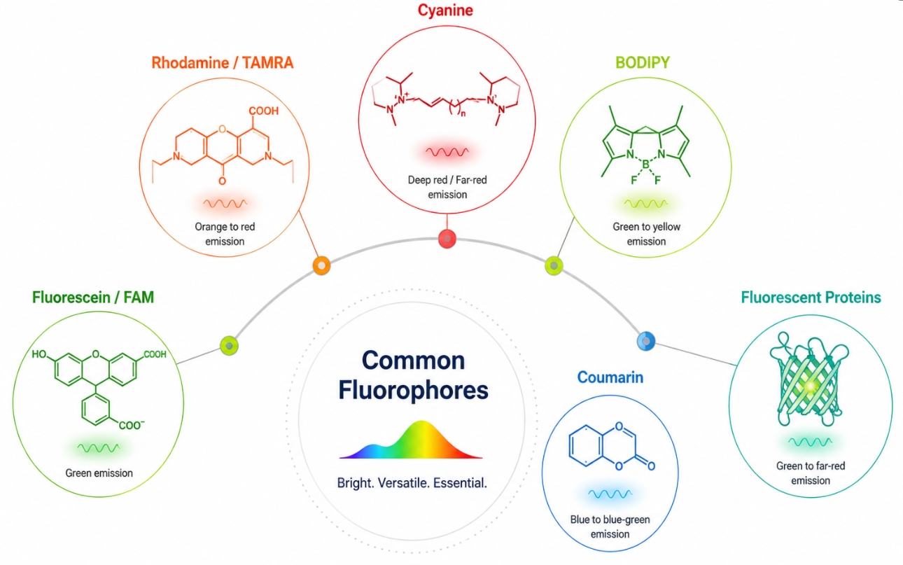 Fig. 1. Several fluorophore families are commonly used in fluorescent labeling (BOC Sciences Authorized).
Fluorescein and FAM-Type Fluorophores
Fluorescein-type fluorophores are among the most familiar labels in fluorescent labeling workflows. They are often used when green-channel detection is suitable and when broad reagent familiarity is an advantage. Their popularity comes from long-standing use in biomolecule labeling, nucleic acid probes, and general fluorescence workflows. They can be highly practical in standard assays, but their selection should still be evaluated carefully because green channels are often crowded, and not every workflow benefits from choosing the most familiar family. In addition, fluorescein-based labels can be very useful in screening and routine fluorescence experiments, yet they may become less ideal in multicolor systems where green-emission overlap, background autofluorescence, or demanding stability requirements start to matter more.
Rhodamine Dyes and TAMRA Fluorophores
Rhodamine-type fluorophores, including TAMRA-related options, are frequently chosen when stronger signal stability and orange-to-red placement are desirable. These fluorophores are common in labeling strategies for proteins, antibodies, probes, and other conjugates where users want a balance between visibility and broader panel flexibility. They often appeal to projects that need a shift away from heavily used green channels while preserving strong detectability and reasonably mature conjugation options. In practical terms, rhodamine-family fluorophores are often attractive when the workflow benefits from more robust imaging performance, clearer channel allocation, or a conjugation route that fits standard biolabeling chemistry without requiring unusually specialized design.
Cyanine Fluorophores
Cyanine fluorophores are widely used across multicolor fluorescent labeling because they cover a broad spectral range and are especially valuable in red and far-red channel planning. Cy3-, Cy5-, and longer-wavelength analogs are commonly selected when users need better separation from blue and green channels or when deeper red-shifted detection is preferred. Their value in practice often lies in spectral placement and multiplex usefulness rather than in generic brightness claims alone. They are particularly helpful in workflows where instrument configuration already supports far-red detection, where autofluorescence in shorter wavelengths is a concern, or where several targets need to be differentiated without collapsing the panel into a narrow spectral region.
BODIPY Fluorophores
BODIPY-based fluorophores are often chosen for their compact structure, strong fluorescence behavior, and versatility in molecular design. They are especially attractive in projects where fluorophore size, tunability, and chemical adaptability matter. In some workflows, BODIPY scaffolds provide a useful balance between strong signal and manageable structural burden, making them relevant not only for general labeling but also for more tailored probe and conjugate development strategies. Their design flexibility can also make them appealing when users need to fine-tune spectral properties, preserve target performance more carefully, or build fluorophore-bearing molecules that must satisfy both optical and structural constraints.
Coumarin, Pyrene, and Other Specialty Fluorophores
Specialty fluorophores such as coumarin- and pyrene dyes-based labels occupy a more selective role in fluorescent labeling. They are often chosen for particular spectral windows, specific scaffold preferences, or specialized assay designs rather than for broad default use. These fluorophores may be especially useful when a less crowded spectral region is needed or when the design constraints of the target favor a more niche fluorophore family. In many cases, their value lies less in broad popularity and more in how well they solve a specific technical problem, such as avoiding common channel congestion, fitting an unusual labeling design, or enabling a probe architecture that more conventional dye families do not support as effectively.
Fluorescent Proteins and Genetically Encoded Fluorophores
Although they are not small-molecule dyes, fluorescent proteins still belong in the broader fluorophore discussion because they represent another major way to generate fluorescence in labeled systems. They are most relevant when the signal is introduced through expression rather than post-labeling conjugation. Their selection logic differs from that of conventional dyes, but they still compete conceptually for spectral space, brightness requirements, imaging duration, and multicolor planning decisions in many fluorescence-based experiments. They are especially important in live-cell observation and genetically encoded workflows where the fluorophore must remain compatible with expression dynamics, protein folding, maturation time, and localization fidelity in addition to ordinary optical criteria.
What Makes a Good Fluorophore?
A good fluorophore is not defined by one parameter alone. In practice, fluorophore performance emerges from a combination of optical properties, chemical behavior, labeling compatibility, and workflow fit. The most reliable way to evaluate a fluorophore is therefore to compare the main selection factors in a structured way rather than prioritizing a single headline attribute such as brightness or wavelength. The table below summarizes the major factors that commonly shape fluorophore choice in fluorescent labeling experiments.
| Selection Factor | What It Means | Why It Matters in Fluorescent Labeling |
|---|---|---|
| Brightness | Overall apparent signal strength produced by the fluorophore. | Influences how easily the labeled target can be detected, especially in low-abundance or low-sensitivity workflows. |
| Quantum Yield | Efficiency with which absorbed energy is converted into emitted fluorescence. | Affects how much useful signal is generated after excitation. |
| Extinction Coefficient | How strongly the fluorophore absorbs excitation light. | Contributes to apparent brightness and helps determine whether excitation is efficient on the chosen platform. |
| Photostability | Resistance to signal loss during repeated or prolonged illumination. | Critical for time-lapse imaging, repeated scanning, and long exposure workflows. |
| Excitation Wavelength | The light range needed to excite the fluorophore efficiently. | Must match available lasers, lamps, or LED sources to avoid weak excitation. |
| Emission Wavelength | The spectral region where fluorescence is emitted. | Determines detector fit, channel placement, and whether the signal can be collected cleanly. |
| Stokes Shift | The separation between excitation and emission peaks. | Larger separation can simplify detection and reduce excitation bleed-through in some workflows. |
| Spectral Overlap | The extent to which one fluorophore's emission overlaps another channel. | High overlap complicates multicolor analysis and increases compensation or separation burden. |
| Solubility | How readily the fluorophore remains dissolved under labeling conditions. | Poor solubility can reduce usable concentration and create inconsistent labeling performance. |
| Hydrophobicity | The tendency of the fluorophore to prefer nonpolar environments. | High hydrophobicity can increase nonspecific binding, aggregation, or target disturbance. |
| Charge | The net electrostatic character of the fluorophore or conjugate. | Can influence solubility, target behavior, cellular distribution, and background signal. |
| Aggregation Tendency | The likelihood that fluorophore molecules cluster or self-associate. | Aggregation can weaken signal quality, complicate interpretation, and alter target behavior. |
| pH Sensitivity | The extent to which fluorescence changes across pH conditions. | Important when the sample environment or assay conditions are not pH-neutral. |
| Environmental Sensitivity | Changes in fluorescence based on polarity, binding context, or local surroundings. | Can be useful in some designs but can also complicate quantitative interpretation if not expected. |
| Reactive Group Availability | Whether the fluorophore is available with useful conjugation handles. | Directly affects how easily it can be attached through NHS esters, maleimides, azides, alkynes, or related chemistries. |
| Labeling Efficiency | How effectively the fluorophore can be incorporated into the target system. | Poor labeling efficiency can leave signal too weak or create inconsistent conjugate populations. |
| Target Compatibility | How well the fluorophore fits the size, chemistry, and sensitivity of the labeled target. | Especially important for peptides, small molecules, and function-sensitive biomolecules. |
| Background Tendency | The likelihood of nonspecific signal or off-target accumulation. | High background can reduce effective contrast even when nominal brightness is high. |
| Multiplex Suitability | How well the fluorophore can coexist with other channels. | Important in panel design, channel separation, and interpretation of multicolor data. |
| Platform Fit | Whether the fluorophore matches the detection hardware and assay format. | A fluorophore that fits one platform well may perform poorly on another. |
| Storage and Handling Stability | How well the fluorophore tolerates storage, solution preparation, and repeated use. | Affects reagent reliability and practical repeatability in real laboratory workflows. |
How to Match a Fluorophore to Your Instrument?
Instrument fit is one of the most practical filters in fluorophore selection because even a chemically suitable label can underperform if the optical platform cannot excite it efficiently or detect it cleanly. From a development perspective, this section is not just about checking whether a fluorophore "works" on a microscope or flow cytometer. It is about reducing avoidable redesign later in the project. A fluorophore should be selected with the real instrument configuration, channel architecture, acquisition mode, and future assay expansion needs in mind. This is especially important for customers building workflows that need to remain stable across optimization, scale-up, multicolor integration, or repeated use on shared instruments.
Match Excitation and Detection Windows First
The first technical question is whether the fluorophore can be excited efficiently and detected within the actual hardware limits of the instrument. In development work, this means checking more than the fluorophore's nominal excitation and emission peaks. The usable excitation range must be matched against available lasers, lamps, or LED sources, while the emission profile must fit the instrument's detector windows and filter sets with enough margin to generate a strong and separable signal. A fluorophore that looks acceptable on a spectrum plot may still underperform if excitation is suboptimal or if the detector only captures part of the emitted signal. For customers, this step is important because it helps avoid investing in a labeling route that later appears weak not because of poor chemistry, but because the fluorophore was never well aligned with the platform in the first place.
Plan for Channel Separation and Panel Expansion
Instrument matching becomes more demanding when the workflow includes more than one fluorescent channel. In multicolor development, the fluorophore should not be judged only by whether it is visible on its own, but by how cleanly it coexists with the rest of the panel. Spectral spillover, compensation burden, and channel crowding can all reduce the practical value of an otherwise attractive fluorophore. From a customer need perspective, this matters because many projects do not stay single-color for long. A fluorophore selected for an early-stage proof-of-concept may later need to coexist with nuclear stains, phenotypic markers, structural stains, or assay-specific reporters. Choosing a channel that leaves room for future expansion can reduce later revalidation work and make the overall labeling strategy more durable as the project evolves.
Align Fluorophore Performance with Acquisition Conditions
Instrument compatibility also depends on how the signal will be acquired, not only on the optical layout of the platform. Long exposures, repeated scanning, time-lapse imaging, fast acquisition, and low-signal detection all place different demands on fluorophore behavior. In development settings, this means the fluorophore should be evaluated against the real observation pattern rather than against a single static image or endpoint readout. A dye that is sufficiently bright in a brief test exposure may become less suitable when repeated illumination accelerates bleaching or when rapid acquisition requires stronger instantaneous signal. For customers building robust workflows, this is a key development consideration because acquisition style often changes as the assay matures. A fluorophore that remains reliable across realistic acquisition conditions is usually more valuable than one that performs well only under ideal short-exposure testing.
Choose for the Platform You Need to Support
Different fluorescence platforms reward different fluorophore characteristics, so instrument matching should always be platform-specific. In fluorescence microscopy, spatial contrast, photostability, and image clarity are often central. In flow cytometry, laser compatibility, detector alignment, and clean channel separation may be more important than image-based contrast. In broader analytical or screening workflows, such as plate-reader or high-throughput formats, uniform detectability and signal robustness across many samples may become the priority. From a customer and development standpoint, this means fluorophore selection should be tied to the platform that will ultimately define success. If the same labeled reagent may later be used across several platforms, the fluorophore should be chosen with cross-platform compromise in mind, so that early optimization does not lock the project into a label that only performs well in one narrow readout mode.
How to Choose by Target and Labeling Chemistry?
Fluorophore selection cannot be separated from what is being labeled and how the fluorophore will be attached. A fluorophore that works well in one target class may behave poorly in another because of size, charge, hydrophobicity, or conjugation requirements. This is especially important when the labeled target must remain functional after modification. In many projects, the most useful fluorophore is not the optically strongest option but the one that best preserves the behavior of the target while still producing interpretable fluorescence.
Protein Labeling Needs
Protein labeling usually requires balancing signal output with preservation of higher-order structure, binding behavior, solubility, and biological activity. Many proteins present multiple accessible lysines or cysteines, which makes labeling chemically feasible but can also introduce heterogeneity in fluorophore placement and labeling degree. From a technical standpoint, the fluorophore should be selected not only for optical performance but also for how much structural burden it introduces after conjugation. Highly hydrophobic or bulky fluorophores can increase aggregation risk, alter folding stability, or disturb interaction surfaces that are functionally important. This is especially relevant for enzymes, receptors, carrier proteins, and interaction-sensitive biomolecules where even moderate over-labeling can shift activity or binding profiles. For protein projects, a good fluorophore is therefore one that provides sufficient signal while keeping conjugation density, steric interference, and physicochemical disturbance within an acceptable range.
Antibody Labeling Needs
Antibody labeling has stricter functional constraints than many general protein-labeling workflows because the fluorescent conjugate is often expected to preserve high target specificity while remaining clean in complex staining environments. The fluorophore must therefore be chosen with attention to antigen-binding preservation, background behavior, and assay architecture. If the label is too hydrophobic, too strongly self-associating, or introduced at excessive density, it can increase nonspecific interactions or reduce effective binding performance. In direct antibody labeling, the fluorophore also becomes part of the detection reagent itself, which means its brightness, stability, and spectral placement directly influence assay sensitivity and multiplex compatibility. In practice, fluorophore choice for antibodies should be judged by how well it supports both recognition fidelity and signal clarity under real staining conditions rather than by brightness alone.
Peptide Labeling Needs
Peptide labeling is often more technically sensitive than protein labeling because the fluorophore can account for a much larger fraction of the final conjugate's size, polarity, and steric profile. As a result, fluorophore choice can significantly alter binding, membrane interaction, conformational behavior, or uptake properties. This makes compactness, linker design, and attachment site more critical than they might be in larger biomolecules. A fluorophore that performs well on a stable protein scaffold may substantially distort the function of a short targeting peptide or signaling peptide. For peptide workflows, the goal is usually not to maximize signal at any cost, but to preserve the functional identity of the peptide while still generating enough fluorescence for reliable detection. This often means favoring fluorophores with manageable structural burden and carefully considering whether the label will be placed at the N-terminus, C-terminus, or a designated side-chain handle.
Nucleic Acid Labeling Needs
Nucleic acid labeling follows a different design logic because fluorophore behavior is closely tied to sequence context, probe architecture, and readout mechanism. The placement of the fluorophore can influence hybridization efficiency, local quenching, spacing relative to complementary strands, and the performance of amplification- or probe-based detection systems. In these projects, fluorophore selection should account not only for excitation and emission properties but also for how the label fits within the broader detection design. A fluorophore that is chemically easy to install may still be suboptimal if it reduces probe discrimination, affects nucleic acid folding, or generates less predictable signal in hybridization-based assays. For customers developing labeled oligonucleotides, primers, or fluorescent probes, the most suitable fluorophore is often the one that supports assay architecture cleanly rather than the one with the most attractive standalone optical profile.
Small-Molecule and Ligand Labeling Needs
Small-molecule and ligand labeling is especially demanding because the fluorophore can dramatically change molecular behavior even when the conjugation itself is chemically straightforward. Parameters such as molecular size, charge, hydrophobicity, and linker position can alter permeability, distribution, receptor affinity, off-target interactions, or intracellular localization. In this setting, fluorophore choice becomes part of the molecular design problem rather than a simple reporting decision. A dye that is acceptable for protein conjugation may be far too disruptive for a low-molecular-weight ligand or inhibitor. This is why small-molecule labeling usually requires a more conservative and structure-aware approach, with careful attention to whether the fluorescent analog still reflects the parent compound's biological behavior closely enough to support meaningful interpretation.
Choosing a Fluorophore for a New Labeling Workflow?
We can help match fluorophore properties, reactive chemistry, and instrument requirements to your experimental goals.
How to Choose for Different Experimental Goals?
Fluorophore selection changes with the experimental goal because not every workflow values the same performance dimensions. Some projects prioritize image contrast and structural readability. Others depend on panel architecture, rapid acquisition, or repeated observation over time. A fluorophore that is excellent for one purpose may be less suitable for another, even when the target is the same. Defining the experimental goal clearly helps narrow the selection to fluorophores whose strengths actually support the intended readout.
Fluorophores for Cellular Imaging
Cellular imaging workflows usually place a premium on spatial contrast, signal localization, and the ability to distinguish the labeled target from surrounding structures or background autofluorescence. In this context, fluorophore choice should be guided not only by nominal brightness but also by how cleanly the signal can be resolved within the cellular environment. A fluorophore with acceptable spectral placement but poor photostability or a tendency toward nonspecific accumulation can reduce image interpretability even when the initial signal appears strong. For imaging-driven projects, the best fluorophore is typically one that combines sufficient brightness, manageable background, and optical clarity under the actual acquisition settings rather than under idealized single-frame conditions.
Fluorophores for Live-Cell Observation
Live-cell workflows impose additional constraints because the fluorophore must remain useful under repeated illumination while minimizing avoidable disturbance to the biological system. Photostability, cellular compatibility, and signal consistency across time become especially important when the experiment involves dynamic localization, trafficking, redistribution, or time-dependent response analysis. In these settings, a fluorophore that is very bright at the beginning but rapidly bleaches or alters target behavior can be less valuable than a moderately bright fluorophore that remains stable and less disruptive over the real observation window. From a technical standpoint, live-cell selection should always consider the combined effect of fluorophore chemistry, illumination burden, and target sensitivity rather than relying on signal intensity alone.
Fluorophores for Flow Analysis
Flow-based assays prioritize different performance factors from microscopy-based workflows. In flow cytometry, fluorophore choice is strongly shaped by laser compatibility, detector alignment, spectral spillover, and overall panel balance. The practical objective is not simply to maximize fluorescence from one marker, but to ensure that each fluorophore contributes a distinguishable signal without creating unnecessary compensation burden or reducing the interpretability of neighboring channels. This means that channel placement and spectral separation often matter more than raw brightness. For customers developing multicolor flow panels, the most useful fluorophore is usually the one that fits cleanly within the panel structure and preserves resolution across the full marker set.
Fluorophores for Immunofluorescence Workflows
Immunofluorescence workflows require fluorophores that perform reliably within antibody-based staining systems, where background control, signal specificity, and multichannel readability are all important. Because the fluorophore is part of a broader staining architecture rather than an isolated label, its usefulness depends on how well it integrates with fixation conditions, antibody behavior, and other markers in the same experiment. A fluorophore that is too spectrally crowded, too background-prone, or insufficiently stable under image acquisition can complicate interpretation even if antibody binding is strong. For immunofluorescence applications, fluorophore choice should therefore support not only visibility, but also cleaner panel structure, lower nonspecific signal, and more robust image interpretation under realistic staining conditions.
Fluorophores for Nucleic Acid Detection
Nucleic acid detection workflows place more emphasis on probe architecture, fluorophore placement, and signal discrimination than many general labeling applications. In these systems, the fluorophore often participates directly in the logic of the assay, whether through hybridization-based readout, probe separation, or amplification-linked detection. Selection should therefore account for more than simple optical brightness. A fluorophore may need to remain compatible with oligonucleotide synthesis, sequence-specific placement, quenching design, or downstream analytical interpretation. From a development perspective, the optimal fluorophore in nucleic acid workflows is often the one that supports clean probe performance and stable assay behavior rather than the one with the strongest standalone fluorescence profile.
Common Fluorophore Selection Errors That Weaken Labeling Results
Many fluorophore selection problems are avoidable, but they persist because the wrong criteria are often used too early. Researchers may choose by color family, brightness reputation, or catalog familiarity before considering how the fluorophore fits the instrument, the target, and the assay structure. These mistakes do not always eliminate fluorescence. More often, they create weak contrast, complex compensation burdens, target disturbance, or analytical ambiguity that only becomes obvious after significant time has already been invested.
- Choosing by Color Name Alone: Selecting a fluorophore because it is simply "green," "red," or "far-red" is too coarse for most modern fluorescent labeling workflows. Color names do not capture excitation efficiency, emission boundaries, detector fit, or how the fluorophore behaves relative to the rest of the panel. Two fluorophores that sound similar in casual language may perform very differently on the same instrument. A useful selection process therefore has to move beyond color labels and into real spectral and workflow comparisons.
- Prioritizing Brightness Without Context: Brightness is useful, but overvaluing it can lead to poor choices. A very bright fluorophore can still create problems if it photobleaches quickly, overlaps heavily with neighboring channels, or disturbs the target after conjugation. Brightness should be evaluated in relation to the platform, the target, and the rest of the workflow rather than treated as a universal ranking criterion. In many experiments, the most successful fluorophore is not the brightest one, but the one that stays interpretable under the full experimental design.
- Ignoring Spectral Spillover: Spectral spillover is one of the most common causes of multicolor interpretation problems. A fluorophore may seem acceptable in single-channel planning but become difficult to separate once additional labels are present. Ignoring overlap early can force more aggressive compensation later and reduce confidence in the final dataset. It can also limit the addition of future channels, making the panel harder to expand or adapt as the experiment develops.
- Overlooking Target Disturbance: Fluorophores are not neutral decorations. They can change hydrophobicity, charge distribution, steric profile, and interaction behavior, especially in smaller or more sensitive targets. If target disturbance is ignored during selection, the resulting labeled molecule may no longer reflect the original biological system accurately even if the fluorescence looks strong. This problem is particularly important in peptide, ligand, and small-molecule labeling, where the fluorophore can have a disproportionately large effect on final behavior.
- Treating Reactive Chemistry as an Afterthought: A fluorophore cannot be chosen independently of its usable reactive forms. If the selected fluorophore is poorly matched to the required labeling chemistry, the project may end up with inefficient conjugation, difficult purification, or a need to redesign the entire route. Chemistry and fluorophore family should be considered together from the beginning. A fluorophore that is excellent optically but awkward chemically may slow development more than a slightly less ideal dye that fits the labeling route cleanly.
- Skipping Workflow-Level Validation: Another common mistake is assuming that acceptable catalog specifications guarantee acceptable experimental performance. In reality, fluorophore selection should always be checked against the real workflow: platform fit, target sensitivity, multicolor compatibility, background behavior, and downstream interpretation. Without this broader validation logic, fluorophore choice remains only partially informed. Validation at the workflow level helps confirm that the fluorophore does not only work in principle, but works in the actual experiment being designed.
A Practical Fluorophore Selection Workflow
In real fluorescent labeling projects, fluorophore selection works best when it follows a structured narrowing process rather than an open-ended comparison of all available dyes. A stepwise approach reduces avoidable trial and error, keeps chemistry and platform planning aligned, and improves the chances that the final label will remain useful beyond the first successful signal. The process below provides a practical way to move from broad possibilities to a shortlist that is technically and analytically credible.
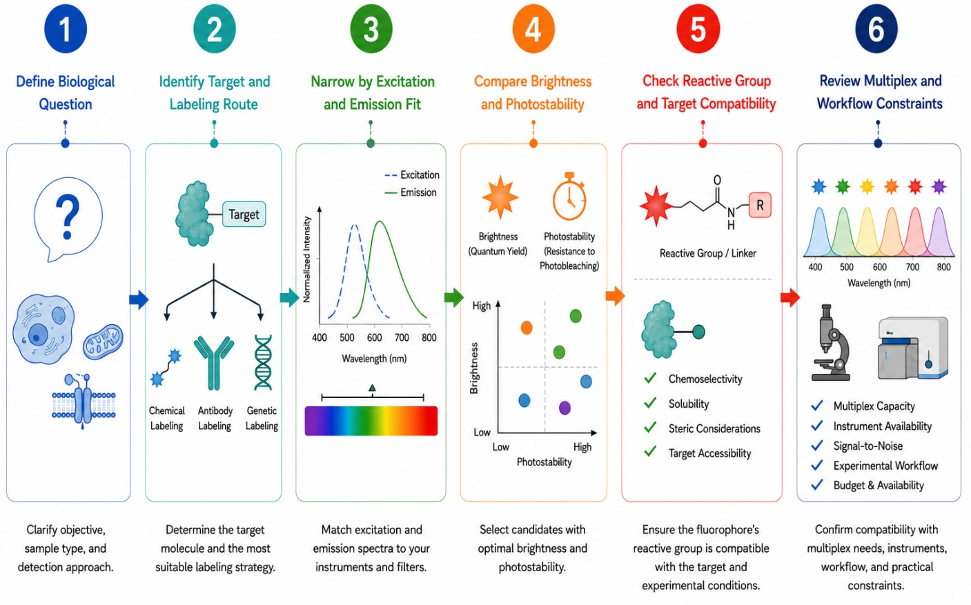 Fig. 2. A structured workflow helps narrow fluorophore choices more efficiently (BOC Sciences Authorized).
- 1. Define the biological question and readout first. Begin by clarifying what the experiment is actually meant to show. Decide whether the goal is localization, tracking, quantification, binding analysis, multicolor panel integration, or repeated observation over time. Also determine whether the system is live or fixed and whether the signal needs to support one endpoint or many observations. This first step matters because it sets the criteria by which every later fluorophore choice will be judged.
- 2. Identify the target type and labeling route. Determine whether the project involves proteins, antibodies, peptides, nucleic acids, small molecules, or another target class, and then define the likely labeling route. A fluorophore that is reasonable for antibody labeling may not be equally suitable for a small molecule or peptide. Clarifying the target and attachment logic early prevents later selection from drifting toward fluorophores that are attractive in general but poorly matched to the actual conjugation problem.
- 3. Narrow candidates by excitation and emission fit. Once the biological context is defined, reduce the candidate list according to the instrument. Remove fluorophores that do not fit the available excitation sources, filter sets, emission windows, or panel structure. This step can eliminate many poor candidates very quickly and helps ensure that the remaining fluorophores are at least technically compatible with the real detection platform rather than only with idealized spectral diagrams.
- 4. Filter by brightness, photostability, and background risk. From the instrument-compatible candidates, evaluate which fluorophores are likely to provide enough signal under the actual acquisition conditions. Consider not only nominal brightness but also how the fluorophore behaves during repeated illumination, how likely it is to generate background, and whether its overall signal profile remains useful for the intended experiment. This step helps avoid the common mistake of choosing a dye that looks powerful initially but becomes less practical after illumination or panel integration.
- 5. Check reactive group and target compatibility. Confirm that the fluorophore is available in a usable reactive form and that its structural and chemical properties are acceptable for the chosen target. This means checking whether the fluorophore can be attached through the required chemistry and whether its size, hydrophobicity, charge, or scaffold are likely to disturb target behavior. A good fluorophore candidate at this stage is one that remains plausible both optically and chemically.
- 6. Review multiplex, purification, and workflow constraints. Before final selection, evaluate whether the fluorophore still fits once the full workflow is considered. This includes panel compatibility, cleanup burden, labeling uniformity, target validation requirements, and how easily the final readout can be interpreted. A fluorophore that survives this final review is more likely to remain useful across real experimental conditions rather than only in preliminary planning.
How BOC Sciences Supports Fluorophore Selection and Labeling Development?
Fluorophore selection is often where fluorescent labeling projects either become more efficient or accumulate avoidable complexity. In many cases, the challenge is not simply identifying a fluorophore that can work, but choosing one that matches the target molecule, conjugation route, platform constraints, and final analytical goal. BOC Sciences supports this process through fluorophore matching, labeling strategy support, conjugation-oriented reagent planning, and development assistance for more specialized fluorescent labeling projects.
Fluorophore Matching for Labeling Projects
- Support for selecting fluorophore families according to target type, spectral range, and downstream experimental purpose rather than relying on color preference alone.
- Comparative guidance across fluorescein, rhodamine, cyanine, BODIPY, coumarin, and related fluorophore classes according to signal, stability, and workflow fit.
- Assistance with narrowing fluorophore options for protein, antibody, nucleic acid, peptide, and small-molecule labeling projects.
- More practical fluorophore selection support for users balancing brightness, stability, and target compatibility in real labeling workflows.
Reactive Group and Conjugation Support
- Guidance on matching fluorophore selection to available reactive forms and conjugation strategies, including amine-reactive, thiol-reactive, and handle-based routes.
- Support for choosing labeling chemistry according to target accessibility, required control level, and downstream purification needs.
- Better coordination between fluorophore family and conjugation route so that signal performance and labeling practicality are considered together.
- Development-oriented support for projects that need more than a standard one-step fluorescent conjugation approach.
Multicolor and Platform Planning
- Assistance with fluorophore selection for fluorescence microscopy, cell imaging, flow-based analysis, and related fluorescence platforms.
- Support for evaluating spectral separation, channel crowding, and signal compatibility in multicolor workflows.
- More structured planning for users who need fluorophores that work cleanly within existing panel or instrument constraints.
- Help with aligning fluorophore properties to acquisition style, observation duration, and downstream interpretation requirements.
Custom Labeling Development Support
- Support for developing fluorescent labeling solutions that require customized fluorophore selection, conjugation logic, or target-oriented optimization.
- Assistance with balancing signal output and target preservation in projects where standard catalog-level decisions are not sufficient.
- Practical development support for users designing fluorescently labeled biomolecules, probes, or functional conjugates with more specific performance goals.
- Project-focused assistance that connects fluorophore choice with the broader demands of labeling development rather than treating dye selection as an isolated step.
Do You Need A Consultation?
BOC Sciences supports fluorophore selection, conjugation planning, and fluorescent labeling development for workflows that require better signal fit, cleaner chemistry, and stronger analytical reliability.
Representative Fluorophores for Labeling Development
| Catalog | Name | CAS | Inquiry |
|---|---|---|---|
| R10-0005 | 6-Fluorescein phosphoramidite | 204697-37-0 | Bulk Inquiry |
| F04-0033 | 5-Aminofluorescein | 3326-34-9 | Bulk Inquiry |
| F01-0012 | 3-Bodipy-propanoic acid | 165599-63-3 | Bulk Inquiry |
| F02-0056 | Gadodiamide | 131410-48-5 | Bulk Inquiry |
| F07-0049 | 6-Carboxytetramethylrhodamine succinimidyl ester | 150810-69-8 | Bulk Inquiry |
| F01-0053 | 8(4'-bromophenyl)-1,3,5,7-tetramethyl-BODIPY | 850534-66-6 | Bulk Inquiry |
| F05-0014 | 7-Hydroxycoumarin-3-carboxylic acid | 779-27-1 | Bulk Inquiry |
| F02-0118 | Cyanine 5 Phosphoramidite | 351186-76-0 | Bulk Inquiry |
| F04-0034 | 5-Carboxyfluorescein diacetate | 79955-27-4 | Bulk Inquiry |
| F04-0055 | Dexamethasone Fluorescein | 216854-76-1 | Bulk Inquiry |
| F05-0031 | 6-Carboxy-X-rhodamine | 194785-18-7 | Bulk Inquiry |
| F06-0006 | 7-Diethylamino-4-methylcoumarin | 91-44-1 | Bulk Inquiry |
| F01-0129 | NIR-BODIPYs-free acid | 1996627-88-3 | Bulk Inquiry |
| F01-0121 | 3-styryl-BODIPYs | 1321616-68-5 | Bulk Inquiry |
| F01-0003 | BDP 558/568 carboxylic acid | 150173-72-1 | Bulk Inquiry |
High-Performance Fluorescent Tools for Your Research
- Cyanine Versatile fluorophores for bioimaging applications.
- JOE Dyes Green fluorescent probes for qPCR applications.
- Coumarin Blue fluorescent probes for enzyme assays.
- TAMRA Dyes Red fluorescent labeling for biomolecule tracking.
- BODIPY Photostable dyes for lipid and cell imaging.
- ICG Dyes Near-infrared imaging and in vivo diagnostics.
- Other Cyanine Tunable near-infrared bioimaging and labeling.
- sulfo-Cyanine Water-soluble cyanine dyes for labeling.
Explore More Topics
Online Inquiry
