

Fluorescent Labeling Methods: Chemical, Immunological, Genetic and Click Approaches
Fluorescent labeling is not a single technique but a family of strategies for introducing a detectable fluorescent signal into a target system. In practice, researchers are often not asking whether a sample can be made fluorescent at all. The more important question is how the signal should be introduced so that it remains specific, interpretable, and compatible with the real workflow. A chemically labeled protein, an antibody-based fluorescent readout, a genetically encoded fluorescent fusion, and a bioorthogonal click-labeled target can all produce useful fluorescence, but they do so through very different logic. Those differences matter because they shape what can be labeled, when fluorescence appears, how much control the user has over signal placement, and how much experimental burden is introduced during preparation and analysis.
This is why method choice deserves to be treated as a first-order design decision rather than a downstream technical detail. In one project, direct chemical conjugation may be the cleanest and fastest route. In another, antibody-mediated recognition may provide the only realistic way to achieve target specificity in a complex sample. In live-cell dynamic observation, fluorescent proteins may offer the continuity that a post-fixation method cannot. In more selective or modular designs, click chemistry reagents or other bioorthogonal strategies may solve problems that traditional amine- or thiol-based conjugation cannot address cleanly. A useful comparison page therefore needs to go beyond naming methods. It has to explain what each route is built to do, where it performs best, where it creates practical trade-offs, and how to compare those trade-offs against the biological question being asked.
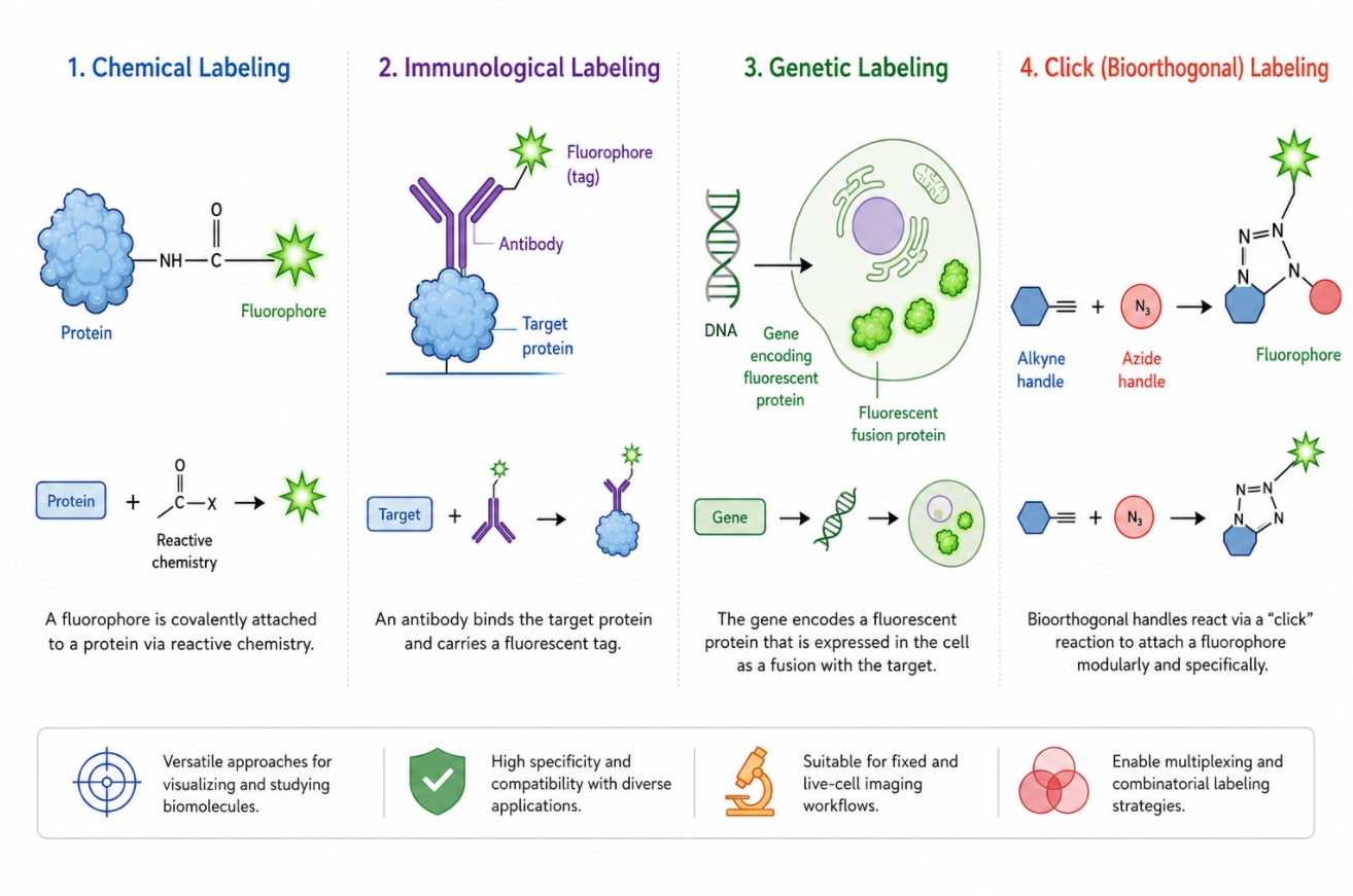 Fig. 1. Four major fluorescent labeling routes differ in how fluorescence is introduced, controlled, and interpreted (BOC Sciences Authorized).
Method selection also sits inside a broader experimental system that usually includes fluorescent dyes, fluorescent probes, sample handling decisions, and one or more downstream detection platforms. A method that looks attractive in isolation may become much less practical once purification, multicolor compatibility, target accessibility, live-cell requirements, or target-function preservation are considered. The purpose of this guide is to compare four major fluorescent labeling routes—chemical, immunological, genetic, and click-based approaches—in a way that helps researchers decide which one fits a real project rather than which one sounds most advanced in principle.
Why Fluorescent Labeling Method Choice Matters
The strongest fluorescent labeling workflow is not necessarily the one that generates the brightest initial image. It is the one that introduces a fluorescent signal in a way that remains analytically meaningful from labeling through readout. Different methods do not merely provide alternative technical flavors of the same operation. They make different assumptions about what the target is, how much specificity is already available, whether the sample is alive or fixed, whether time-resolved observation is needed, and how much modification the target can tolerate. When these assumptions are mismatched to the real experiment, the result is often not total failure but a more subtle loss of interpretability. The signal may still be visible, yet the biology is no longer being reported in the right way.
Different Methods Answer Different Experimental Questions
Each fluorescent labeling route is best understood by the kind of problem it solves. Chemical labeling is often chosen when a defined molecule is already available and the user wants a direct route to a fluorescent conjugate through a known reactive handle. Immunological labeling is most useful when the key challenge is target recognition in a structurally or compositionally complex sample, especially when endogenous targets must be detected without prior modification. Genetic labeling becomes attractive when the goal is continuous signal generation inside living systems, particularly for dynamic localization, trafficking, or time-dependent expression studies. Click and broader bioorthogonal approaches are especially valuable when researchers need to separate target installation from fluorophore attachment, improve selectivity inside complicated environments, or build modular labeling routes that are harder to achieve with direct conjugation alone. The question is therefore not which method is universally best, but which method is best aligned with the target, timing, and analytical purpose of the experiment.
The Cost of Choosing the Wrong Labeling Route
A poor method choice often reveals itself as increased complexity rather than immediate failure. Direct chemical conjugation may produce heterogeneous labeling and compromise activity if used on a target that requires tighter site control. Antibody-based labeling may provide excellent specificity but become impractical in live-cell workflows, inaccessible epitopes, or highly multiplexed panels where background and species cross-reactivity become harder to manage. Genetic labeling can generate elegant live-cell readouts, yet it may introduce expression burden, perturb localization, or fail to reflect endogenous protein behavior if the fusion construct changes the system. Click-based labeling may improve selectivity and modularity, but it can also introduce more steps, more reagent dependencies, and more optimization burden than a project can realistically support. In other words, the wrong fluorescent labeling method rarely wastes only signal. It can distort timing, complicate purification, crowd the panel design, or shift the biological meaning of the final readout.
Chemical Fluorescent Labeling
Chemical fluorescent labeling is the most direct route in many projects because it relies on covalent attachment of a fluorophore to an accessible functional group on the target. This makes it highly flexible across proteins, antibodies, peptides, nucleic acid derivatives, and certain small molecules or modified particles. Its popularity comes from its practical simplicity: if the target can be isolated or at least presented with a suitable reactive site, the fluorophore can often be attached without requiring a recognition partner or expression system. Yet that apparent simplicity should not be mistaken for uniform control. Chemical labeling performance depends heavily on reactive group choice, target composition, reaction environment, and how much heterogeneity can be tolerated in the final conjugate.
Reactive Groups Such as NHS Esters, Maleimides, Azides, and Alkynes
Chemical fluorescent labeling is built around reactive group compatibility, and this is one of the main reasons it remains so flexible across different biomolecules and workflow designs. The reactive group determines not only whether a fluorophore can be attached efficiently, but also how much control the user has over site access, labeling uniformity, downstream purification, and target-function preservation. In practice, these chemistries are not interchangeable. Each one is better understood as a different entry point into fluorescent conjugation, with its own balance of accessibility, selectivity, and workflow complexity.
- NHS Esters: NHS esters are among the most widely used reactive groups in fluorescent labeling because they react efficiently with primary amines, especially lysine residues and N-terminal amines on proteins and antibodies. Their practical advantage is accessibility: many biomolecules already present suitable amine sites, which makes NHS ester chemistry a convenient first option for direct dye conjugation. At the same time, this broad accessibility can reduce site control, since multiple amines may be modified within the same target population.
- Maleimides: Maleimides are commonly selected when thiol-directed labeling is preferred, especially in systems where cysteine residues or engineered sulfhydryl groups provide a more controlled attachment site. Compared with broad amine labeling, maleimide chemistry can improve positional control and reduce labeling heterogeneity when the target is designed or prepared appropriately. This makes it especially useful when structural preservation or conjugate uniformity matters more than maximum site availability.
- Azides: Azides are often used as compact chemical handles in modular fluorescent labeling workflows. Their value is not usually in broad direct reactivity, but in how effectively they support staged labeling designs, especially when fluorescence is introduced through a later click-type reaction. Because azides are small and relatively unobtrusive, they can help preserve target behavior during early preparation stages before the final fluorophore is installed.
- Alkynes: Alkynes serve a similar role to azides in handle-based conjugation strategies and are especially important in click-oriented fluorescent labeling routes. They are useful when users want to separate target modification from fluorophore installation, making the overall workflow more modular and sometimes more selective than classic direct conjugation. In practice, alkynes are most valuable when the project benefits from post-installation flexibility rather than immediate one-step labeling.
When Direct Dye Conjugation Works Best
Direct chemical labeling is strongest when the target has accessible reactive sites, can be presented under reasonably controlled conditions, and does not require an intermediary recognition system to define specificity. This often makes it the most straightforward choice for purified proteins, pre-isolated antibodies, designed peptides, modified oligonucleotides, or reagent development workflows in which a fluorescent conjugate is itself the intended product. It is also useful when the user wants to choose the fluorophore independently of the targeting element, since this gives more control over excitation and emission placement, brightness, or multicolor compatibility. In exploratory reagent preparation, direct conjugation is often preferred because it moves quickly from target to fluorescent material without requiring cell engineering or antibody screening. Its practical strength is therefore speed plus modular fluorophore choice, provided the user can manage the consequences of partial site control and post-reaction cleanup.
Common Limitations in Control, Stoichiometry, and Target Integrity
The main limitations of chemical fluorescent labeling arise from the fact that reactive groups do not inherently understand biological importance. If several accessible lysines or cysteines are present, the fluorophore may label a distribution of sites rather than a single defined position. This can broaden the product population, alter labeling stoichiometry from molecule to molecule, and complicate interpretation when the labeled material is expected to behave uniformly. Over-labeling can reduce solubility, change charge distribution, or interfere with binding and catalytic performance. Under-labeling can leave the signal too weak for reliable downstream use. Small targets such as peptides and certain ligands are particularly sensitive because the fluorophore may represent a large structural burden relative to the target itself. Chemical labeling therefore works best when users treat conjugation not as an automatic reaction but as a controlled balance between signal generation, structural preservation, and acceptable product heterogeneity.
Immunological Fluorescent Labeling
Immunological fluorescent labeling uses antibody recognition as the primary source of specificity. Instead of relying on a reactive site that may appear on many molecules, it identifies the target through an epitope and then couples that recognition to fluorescence. This is why immunological labeling is so widely used in complex biological samples where direct chemical selectivity would be difficult to achieve. It can reveal endogenous proteins in heterogeneous mixtures, structured cells, or tissue-derived material without pre-installing a handle on the target. At the same time, its strengths come with a different set of constraints: antibody access must be physically possible, epitope preservation matters, and the fluorescence signal depends on the architecture of the antibody system rather than only on the fluorophore itself.
Direct vs Indirect Immunolabeling
Direct and indirect immunolabeling rely on the same antibody-based recognition principle, but they differ in how fluorescence is introduced and how that choice affects workflow simplicity, signal strength, background control, and multiplex flexibility. The clearest way to compare them is side by side, because the practical difference is not only in reagent architecture but also in how each format performs under real experimental constraints.
| Comparison Point | Direct Immunolabeling | Indirect Immunolabeling |
|---|---|---|
| Fluorophore Placement | The fluorophore is attached directly to the primary antibody. | The fluorophore is attached to a secondary antibody that detects the unlabeled primary antibody. |
| Workflow Complexity | Usually involves fewer incubation and wash steps, making the workflow simpler and easier to control. | Adds an extra detection layer, so the workflow is more complex and requires additional handling. |
| Signal Intensity | Usually provides a cleaner but less amplified signal. | Often produces stronger fluorescence because multiple secondary antibodies can associate with one primary antibody. |
| Background Risk | Often offers better control over nonspecific background because fewer reagents are involved. | Can increase background if secondary antibody binding is not tightly controlled. |
| Reagent Flexibility | Each primary antibody usually needs to be labeled individually. | One labeled secondary antibody can often be reused with multiple primary antibodies from the same host species. |
| Multiplex Design | Can simplify multiplex workflows by reducing species and secondary-antibody constraints. | Requires more careful species planning to avoid cross-reactivity and channel confusion. |
| Best-Fit Use Cases | Better suited to assays that prioritize workflow simplicity, cleaner architecture, and reduced cross-reactivity. | Better suited to workflows that need stronger signal output and broader reagent reuse. |
In practice, the better option depends on what the experiment values most. If the main goal is a shorter and more controlled workflow, direct immunolabeling is often the better fit. If the project needs stronger signal output or more flexible reuse of antibody reagents, indirect immunolabeling is often the more practical choice.
Where Antibody-Based Specificity Is Strongest
Antibody-based fluorescent labeling is particularly powerful when the main analytical challenge is target recognition inside a complex sample. This includes cellular imaging of endogenous proteins, localization analysis in structured specimens, comparative expression studies, and panel-based assays in which several protein targets must be distinguished within the same biological context. Its value is highest when the target is difficult to isolate chemically, when labeling must occur against a background of many chemically similar species, or when the user needs to preserve the native biological context rather than modify the target in advance. In these settings, the antibody provides the specificity that chemistry alone cannot guarantee. The fluorescent signal then becomes a report of antibody-target recognition rather than simple fluorophore attachment, which is why immunological labeling remains central to many imaging and analysis workflows.
Constraints in Live-Cell Workflows, Epitope Accessibility, and Multiplex Settings
Despite its specificity, immunological fluorescent labeling is not universally transferable across workflow types. Many antibody-based methods are strongest in fixed and permeabilized samples because these conditions improve access to intracellular targets and stabilize structural context. In live-cell workflows, antibody entry and epitope exposure may be limited, making the method more suitable for surface targets than intracellular ones unless specialized delivery strategies are used. Epitope accessibility can also change with fixation, conformation, binding partners, or local crowding, which means lack of signal does not always reflect absence of target. In multiplex settings, the advantages of antibody specificity can be offset by species management, secondary cross-reactivity, channel crowding, and the need to separate several fluorophore-labeled recognition events cleanly. A strong immunological design therefore requires not only a good antibody but also careful thinking about access, background, and panel architecture.
Comparing Fluorescent Labeling Methods for a New Workflow?
We can help evaluate chemical, immunological, genetic, and click approaches based on specificity, live-cell compatibility, and downstream analysis needs.
Genetic Fluorescent Labeling with Fluorescent Proteins
Genetic fluorescent labeling introduces fluorescence at the expression level rather than by post-synthetic attachment. The most familiar version uses a fluorescent protein sequence fused to the coding region of the target protein, allowing the target to be produced as a fluorescent chimera inside the biological system itself. This changes the logic of labeling in a major way. Instead of asking where a fluorophore can be attached after the fact, the experiment is designed so that the system generates the fluorescent target continuously. That makes genetic labeling especially valuable for live-cell studies, temporal observation, and dynamic localization analysis. It also means that signal quality depends on expression control, folding, maturation, and biological compatibility, not only on fluorophore brightness.
Fusion Expression as a Labeling Strategy
A fluorescent fusion construct links the target protein to a fluorescent protein sequence so that both are expressed as one product. In principle this provides a highly elegant labeling route because every correctly expressed target molecule carries the label by design. In practice, however, fusion placement matters. An N-terminal tag may behave differently from a C-terminal tag, and in some targets either position can interfere with localization, interaction partners, membrane insertion, or regulatory control. The linker region can also influence flexibility and steric accessibility. Genetic labeling is therefore not simply a cloning task. It is a structural and functional design choice that requires evaluating whether the fusion remains a trustworthy proxy for the unlabeled target.
Advantages for Live-Cell Dynamic Observation
The greatest strength of genetic fluorescent labeling is continuity. Because the fluorescent signal is produced by the biological system, the user can observe target behavior in living cells without adding exogenous staining reagents at every time point. This makes the method particularly attractive for tracking movement, redistribution, assembly, trafficking, or expression-linked changes over time. It also avoids some of the wash, delivery, and accessibility issues that complicate post-labeling methods. For dynamic studies, this continuity often matters more than absolute brightness. A slightly less intense signal that remains tied to the target throughout the experiment may be more valuable than a bright endpoint label that cannot report time-resolved behavior.
Expression Burden, Maturation Time, and Localization Caveats
Genetic labeling does, however, move complexity upstream. The target must be expressed successfully, the fluorescent tag must fold and mature, and the fusion must not substantially alter biological behavior. Overexpression can distort the system even when the fluorescent signal looks excellent. Some targets may mislocalize, aggregate, or change interaction patterns after fusion. Fluorescent protein maturation is not always instantaneous, which can create a lag between target production and detectable fluorescence. In regulated or endogenous-like studies, these issues can be more important than the appeal of built-in signal generation. Genetic fluorescent labeling is therefore most reliable when the fusion construct is treated as an experimental variable that needs validation, not as a neutral tag assumed to leave the biology unchanged.
Click-Chemistry and Bioorthogonal Labeling
Click-based and more broadly bioorthogonal labeling methods are built around a powerful idea: the fluorophore does not always need to be attached directly in the same step that defines the target. Instead, a small and relatively unobtrusive chemical handle can first be introduced into the system, followed later by a selective reaction that brings in the fluorescent component. This separation of handle installation and signal attachment is what gives click-oriented workflows their modularity. They can be especially helpful when classic broad-access chemistries are too indiscriminate, when timing control matters, or when researchers want to preserve target behavior during the first stage and reveal fluorescence at a later point.
Why Bioorthogonal Chemistry Is Attractive
Bioorthogonal logic is attractive because it creates a labeling reaction that is designed to occur selectively in the presence of many other biological functionalities. This can reduce unwanted modification of non-target molecules, simplify the conceptual separation between targeting and reporting, and support workflows in which the same handle-bearing material can later be paired with different fluorophores. It also allows researchers to treat fluorescence as a modular output rather than as an inseparable property of the initial targeting component. In assays that demand higher control, this modularity is often more valuable than simple convenience because it expands how labeling can be timed, tuned, and adapted.
When Click Approaches Outperform Classic Conjugation
Click-oriented fluorescent labeling often outperforms classic direct conjugation when conventional chemistries lack sufficient selectivity or when the target would benefit from a staged workflow. This includes cases where amine-directed labeling would modify too many sites, where a target is better handled first with a minimal chemical handle, or where late-stage fluorophore installation improves flexibility in assay development. These methods can also be useful when the user wants the option to screen several fluorophores against the same handle-bearing intermediate without re-engineering the targeting element each time. In modular probe construction, selective biomolecule derivatization, and advanced conjugation design, click strategies are often preferred not because they are fashionable but because they solve a real control problem.
Practical Limits in Workflow Complexity, Reagent Choice, and Downstream Handling
The price of that control is additional workflow burden. Click-based labeling usually requires more than one design decision: how the handle will be installed, which reactive pair will be used, how efficiently the reaction will proceed in the intended environment, and whether the final construct remains compatible with purification and downstream analysis. The chemistry may be elegant, but it is not automatically simpler. Each extra step introduces another opportunity for partial conversion, background reactivity, incomplete cleanup, or mismatched reagent behavior. For that reason, click-based fluorescent labeling is most valuable when the extra complexity buys something important—greater selectivity, better modularity, later-stage flexibility, or cleaner target preservation—rather than being adopted simply because it sounds more advanced than classic conjugation.
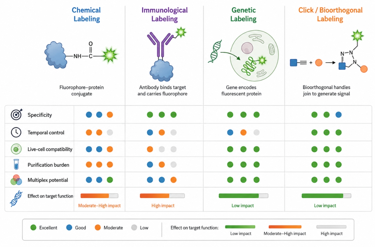 Fig. 2. A practical method comparison matrix helps match labeling routes to specificity, timing, and workflow demands (BOC Sciences Authorized).
How to Compare Fluorescent Labeling Methods for Real Projects
Real method selection works best when it is framed as a decision matrix rather than a popularity contest. No fluorescent labeling route is universally superior across all project types because each one optimizes a different balance of specificity, timing, compatibility, and practical burden. Instead of asking which method is strongest in general, it is more useful to compare how each method behaves under the conditions that actually define the project: Is the sample living or fixed? Does the target already exist in purified form? Is endogenous recognition needed? Must the signal appear continuously or at a chosen time point? Is the panel already crowded? Does the target tolerate chemical modification? The dimensions below provide a practical way to compare methods without reducing them to oversimplified pros-and-cons lists.
| Comparison Dimension | Chemical Labeling | Immunological Labeling | Genetic Labeling | Click/Bioorthogonal Labeling |
|---|---|---|---|---|
| Specificity | Usually depends on reactive group accessibility rather than target recognition, so selectivity can be broad unless the target is well controlled. | Usually strongest in complex samples because specificity comes from antibody-epitope recognition. | Depends on construct design and expression control, with fluorescence genetically tied to the intended target. | Can provide high reaction selectivity by separating handle installation from fluorophore attachment. |
| Temporal Control | Typically introduced as a discrete preparation step before downstream analysis. | Usually applied at a defined staining stage, often after fixation or selected sample preparation steps. | Best suited to continuous signal generation over time inside living systems. | Often supports staged timing by allowing handle introduction first and fluorescent development later. |
| Live-Cell Compatibility | Can be compatible depending on target type, reagent behavior, and recovery requirements. | Often limited for intracellular live-cell targets because access and epitope exposure can be restrictive. | Typically the strongest option for long-duration live-cell observation. | Varies with the exact chemistry and installation route, so compatibility must be judged case by case. |
| Purification Burden | Often requires removal of free dye and management of labeling heterogeneity. | Usually involves wash steps, antibody background control, and panel architecture rather than classic conjugate purification. | Reduces post-labeling cleanup but shifts effort toward construct generation and validation. | Can require both intermediate handling and final cleanup if excess reagent or incomplete conversion would affect analysis. |
| Multiplex Potential | Offers broad fluorophore choice, which is useful in multicolor panel design when site control is acceptable. | Supports multiplexing well, but antibody species, secondary architecture, and spectral crowding must be managed carefully. | Can work well in live multicolor designs if fluorescent proteins are spectrally and biologically compatible. | Often highly modular for multiplex design, though added flexibility comes with more workflow complexity. |
| Effect on Target Function | Can alter charge, hydrophobicity, binding interfaces, or solubility if label load or position is poorly controlled. | Usually does not covalently modify the endogenous target, but antibody binding can still influence accessibility or interpretation. | May change localization, folding, or regulation if the fusion construct perturbs native behavior. | Can reduce some direct-modification risks, but both the installed handle and final fluorophore still need functional evaluation. |
In practical project planning, these criteria are most useful when ranked rather than treated equally. A researcher focused on endogenous protein detection in a structurally complex sample may prioritize specificity and accessibility above all else, which often points toward immunological labeling. A team building a reusable fluorescent conjugate may prioritize direct control over fluorophore family and chemical attachment, which often favors chemical methods. A live-cell dynamics study may prioritize temporal continuity and low intervention during observation, which often makes genetic labeling more attractive. A modular platform that must preserve the target during early stages and reveal fluorescence later may benefit most from click-based design. The correct comparison therefore depends not only on what each method can do, but on which performance dimensions matter most in the intended workflow.
How BOC Sciences Supports Fluorescent Labeling Projects
Fluorescent labeling projects often require more than access to a fluorophore alone. In many cases, the real challenge lies in selecting an appropriate labeling route, matching reactive chemistry to the target molecule, preserving target performance after conjugation, and building a workflow that remains compatible with downstream imaging or analytical readout. BOC Sciences supports fluorescent labeling projects through reagent selection, custom labeling development, fluorophore matching, and method-oriented technical assistance. Our goal is to help researchers move from a general labeling idea to a more practical and better-controlled fluorescent labeling solution.
Fluorescent Labeling Strategy Design
- Support for selecting suitable fluorescent labeling routes according to target type, including proteins, antibodies, peptides, nucleic acids, and other functional molecules.
- Comparative guidance on chemical conjugation, immunological labeling, genetic labeling, and click-based labeling strategies based on specificity, workflow complexity, and downstream assay needs.
- Assistance with evaluating whether direct fluorophore attachment, handle-based labeling, or recognition-driven labeling is the more practical choice for a given project.
- Project-oriented method planning that helps connect biological questions with feasible fluorescent labeling designs rather than treating all routes as interchangeable.
Custom Fluorescent Conjugation and Labeling Development
- Support for developing custom fluorescently labeled biomolecules and related conjugates according to target structure, accessible functional groups, and intended application requirements.
- Flexible development options for amine-reactive, thiol-reactive, and click-compatible fluorescent conjugation workflows where greater control over labeling structure is needed.
- Assistance with fluorophore incorporation strategies designed to balance signal output with target integrity, solubility, and functional preservation.
- Development support for projects that require more specialized labeling architectures, including modular or staged labeling designs rather than only one-step conjugation.
Fluorophore and Reactive Group Matching
- Support for matching fluorophore families, spectral regions, and reactive groups to the chemistry and analytical needs of the target system.
- Guidance on selecting among NHS ester, maleimide, azide, alkyne, and other compatible labeling chemistries according to the desired balance of accessibility, control, and workflow simplicity.
- Assistance with multicolor planning so that fluorescent labels can be integrated more cleanly into microscopy, flow-based, or broader fluorescence analysis workflows.
- Better alignment between fluorophore properties and project requirements such as brightness, spectral separation, purification burden, and signal interpretability.
Technical Support for Fluorescent Labeling Development
- Technical assistance with fluorescent labeling workflow design, including route comparison, reagent matching, and considerations related to labeling efficiency and downstream compatibility.
- Support for reducing common development risks such as excessive modification, poor labeling uniformity, unsuitable fluorophore placement, or avoidable background complications.
- Guidance for adapting fluorescent labeling design to the practical demands of purification, validation, multicolor use, and assay-specific interpretation.
- More structured project support for users developing fluorescent labeling strategies that must be technically credible, application-relevant, and scalable beyond an initial concept stage.
Do You Need A Consultation?
BOC Sciences helps researchers compare fluorescent labeling routes, select compatible reagents, and build workflows that match target biology, assay timing, and downstream analysis requirements.
Reagents Relevant to Fluorescent Labeling Method Development
| Catalog | Name | CAS | Inquiry |
|---|---|---|---|
| F02-0026 | Cy5-NHS ester | 146368-14-1 | Bulk Inquiry |
| F08-0015 | N-(1-Pyrenyl)maleimide | 42189-56-0 | Bulk Inquiry |
| R01-0472 | Atto 425-NHS ester | 892156-28-4 | Bulk Inquiry |
| R01-0005 | BDP 558/568 NHS ester | 150173-73-2 | Bulk Inquiry |
| F04-0027 | Fluorescein-5-maleimide | 75350-46-8 | Bulk Inquiry |
| F03-0035 | sulfoCyanine 3 NHS Ester | 1424433-17-9 | Bulk Inquiry |
| F01-0017 | BDP R6G maleimide | 2183473-32-5 | Bulk Inquiry |
| F01-0228 | BDP 581/591 maleimide | 2183473-29-0 | Bulk Inquiry |
| F01-0006 | BDP 630/650 azide | 2183473-22-3 | Bulk Inquiry |
| F01-0011 | BDP FL azide | 1379771-95-5 | Bulk Inquiry |
| F01-0230 | BDP 581/591 azide | 2183473-20-1 | Bulk Inquiry |
| F07-0015 | TAMRA azide, 5-isomer | 825651-66-9 | Bulk Inquiry |
High-Performance Fluorescent Tools for Your Research
- Cyanine7.5 Extended NIR imaging for in vivo studies.
- JOE Dyes Green fluorescent probes for qPCR applications.
- Fluorescent Dyes General-purpose labeling for bioanalytical detection.
- Cyanine5 Red fluorescence for protein and DNA labeling.
- Cyanine7 Near-infrared probes for optical imaging.
- sulfo-Cyanine Water-soluble cyanine dyes for labeling.
- Other Cyanine Tunable near-infrared bioimaging and labeling.
- Cyanine5.5 Far-red fluorescence for deep tissue imaging.
Explore More Topics
Online Inquiry
