

Fluorescent Labeling vs Fluorescent Probes vs Fluorescent Staining: What Is the Difference?
In fluorescence-based research, the terms fluorescent labeling, fluorescent probes, and fluorescent staining are often used as if they mean the same thing. In practice, they overlap, but they do not describe the same technical role. All three can generate a fluorescent signal, yet they differ in what is being modified, how the signal is introduced, what kind of information the signal is expected to report, and how much control the user has over target definition and workflow design. That difference matters because many experimental errors begin not with poor reagent quality, but with choosing the wrong fluorescence strategy for the biological question.
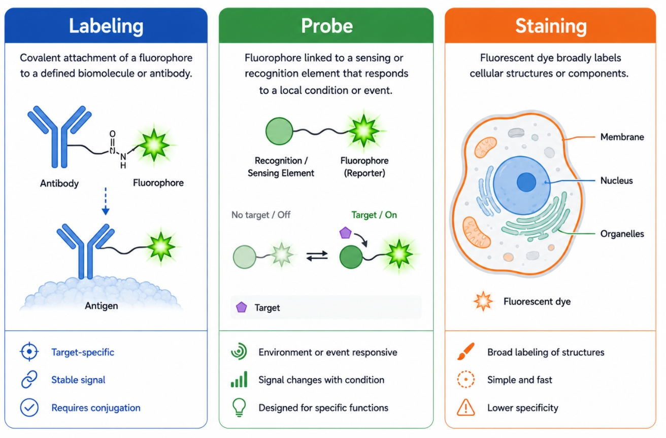 Fig. 1. Fluorescent labeling, probes, and staining serve different technical purposes (BOC Sciences Authorized).
A useful way to think about the distinction is this: fluorescent labeling usually refers to the broader act of attaching or introducing a fluorescent element into a defined target system; fluorescent probes are more often functional molecular tools that recognize, respond to, or report on a target or condition; fluorescent staining is more often a practical visualization approach used to make structures, compartments, or sample classes visible. These categories can overlap in real workflows, and some products may reasonably sit between two of them, but the concepts are still worth separating. Once those boundaries are clear, it becomes much easier to choose the right chemistry, the right fluorophore, the right readout strategy, and the right development path for a given project.
Why These Fluorescence Terms Are Often Confused?
These terms are often confused because fluorescent labeling, fluorescent probes, and fluorescent staining can all produce a similar endpoint: a visible fluorescent signal in an image, assay, or analytical readout. However, they differ in technical purpose and in how that signal is generated. Fluorescent labeling usually emphasizes the deliberate introduction of fluorescence into a defined target through a chosen labeling route. Fluorescent probes more often emphasize recognition, response, or reporting logic, meaning the fluorescence is tied to a molecular event, local condition, or specific type of interaction. Fluorescent staining usually emphasizes practical visualization of structures, compartments, or sample components through binding, partitioning, accumulation, or class-level affinity. In real workflows, these categories can overlap, and product naming does not always preserve strict boundaries, which is why users often encounter the same reagent described with different language across catalogs, protocols, and application notes.
What Fluorescent Labeling Usually Means?
Fluorescent labeling is best understood as the broader technical act of introducing fluorescence into a defined target system. The target might be a protein, antibody, peptide, nucleic acid, small molecule, nanoparticle, cell population, or engineered biological structure. The key feature is not simply that fluorescence becomes visible, but that the fluorescence is intentionally linked to a target through a specific labeling route. In this sense, labeling is a process category rather than a single reagent category. It covers many ways of attaching or installing fluorescence, including direct dye conjugation, recognition-based fluorescent detection, genetically encoded fluorescence, and click-enabled or bioorthogonal strategies.
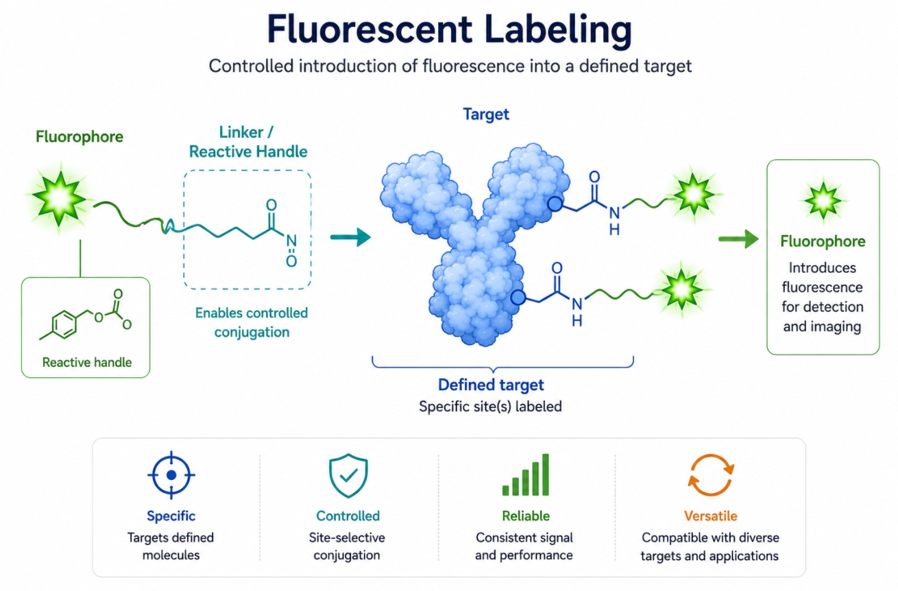 Fig. 2. Fluorescent labeling introduces fluorescence into a defined target through a controlled route (BOC Sciences Authorized).
Fluorescent Labeling as a Target-Directed Process
The defining logic of fluorescent labeling is target direction. The fluorophore is not added merely to brighten the sample in a general way. It is introduced so that fluorescence becomes associated with a selected molecule, structure, or engineered system through a technically controlled route. This makes labeling especially important in workflows where the user needs stronger interpretive confidence about what the signal represents and where that signal originates within the target system.
- Defined Target Association: In labeling workflows, fluorescence is expected to remain connected to a specific target rather than distribute broadly across similar structures or components. This target association may be achieved through covalent conjugation, affinity recognition, genetic fusion, or staged bioorthogonal installation, but in all cases the signal is intended to travel with the selected target rather than with an entire class of sample material.
- Method-Dependent Specificity: The specificity of labeling often comes from the route itself, such as conjugation chemistry, antibody recognition, genetic fusion, or controlled target installation. This means the reliability of the final signal depends not only on fluorophore brightness, but also on reaction selectivity, target accessibility, labeling stoichiometry, and how well the method preserves target identity after modification.
- Higher Structural Control: Compared with general staining, labeling usually places more emphasis on where fluorescence is introduced and how much modification the target can tolerate. This is especially relevant in proteins, antibodies, peptides, or ligands, where excessive labeling can alter folding, binding, solubility, or biological behavior. A good labeling workflow therefore balances signal intensity with structural preservation and interpretability.
- Better Fit for Construct and Assay Development: When the project involves making a reusable fluorescent conjugate or designing a controlled target-tracking workflow, labeling is often the most appropriate conceptual frame. From a development standpoint, labeling is valuable because it creates a clearer relationship between reagent design, target definition, and downstream signal behavior, which is harder to guarantee with more general fluorescence approaches.
Common Fluorescent Labeling Routes
One reason the term fluorescent labeling remains broad is that it includes several technically distinct approaches. These routes differ in how fluorescence is installed, where selectivity comes from, and how much flexibility the user has over timing, stoichiometry, and downstream workflow design. Understanding these routes is important because the meaning of "labeling" changes depending on whether the fluorophore is introduced chemically, biologically, or through a recognition system.
- Chemical Labeling: Fluorophores are attached through reactive groups such as NHS esters or maleimides, often to proteins, antibodies, peptides, or other derivatizable targets. This route is powerful because it can be direct and versatile, but it also requires attention to accessible functional groups, conjugation density, hydrolysis sensitivity, purification burden, and the possibility of heterogeneous labeling populations.
- Immunological Labeling: Fluorescence is introduced through antibody recognition, either by using labeled primary antibodies or by detecting unlabeled primaries with fluorescent secondary reagents. Here, the specificity comes from antibody-target binding rather than from direct target derivatization, which makes the workflow highly useful for endogenous targets but also dependent on antibody quality, background control, species compatibility, and assay architecture.
- Genetic Labeling: Fluorescent proteins or encoded tags are introduced at the expression level so that the target carries fluorescence as part of its produced form. This route can be especially useful for dynamic observation and lineage-linked fluorescence, but it also introduces considerations such as expression burden, folding behavior, tag placement, maturation time, and the possibility that fusion architecture may alter the biology being studied.
- Click or Bioorthogonal Labeling: A handle is installed first and the fluorophore is attached later through a staged, more selective ligation route. This route is attractive when users need more selectivity, lower target perturbation, or more freedom to choose the fluorophore after the target-bearing intermediate has already been prepared. Its strength lies in modularity, but it also introduces additional workflow design and validation requirements.
What Fluorescent Labeling Is Best Suited For
Fluorescent labeling is usually the best framing when the workflow begins with a defined target and the user wants fluorescence to remain meaningfully linked to that target throughout analysis. It is especially useful when reagent architecture, fluorescent identity, and target association all need to be controlled rather than inferred. In these settings, labeling is not simply a visualization tool but part of the analytical design itself.
- Biomolecule Visualization: Useful when the goal is to visualize a known protein, antibody, nucleic acid, ligand, or engineered construct. In these cases, the fluorescence should reflect the selected biomolecule rather than a broad structural category.
- Tracking and Localization: Appropriate when fluorescence needs to follow the behavior, location, or fate of a selected target over time. This is especially important in workflows where movement, compartment transition, or retention needs to be interpreted in relation to one defined labeled entity.
- Conjugate Development: Important in workflows that require a stable fluorescent conjugate rather than a transient staining step. This includes custom fluorescent biomolecules, detection reagents, and assay-specific labeled constructs that must remain reproducible across batches or projects.
- Controlled Assay Design: Valuable when the project requires tight alignment between target identity, fluorophore choice, and downstream detection logic. In these cases, labeling provides a stronger framework for validation, optimization, and troubleshooting because the signal is designed into the system rather than added only for visualization convenience.
What Fluorescent Probes Usually Mean?
Fluorescent probes usually refer to a more function-oriented class of reagents. A probe is not defined only by carrying a fluorophore, but by how that fluorophore is coupled to recognition, response, or reporting behavior. In other words, probes are often designed to do more than make a target visible. They are often meant to detect, bind, sense, respond to, or reveal a specific biological or chemical condition. That makes probes particularly valuable when the user wants information beyond simple target presence, such as local environment, molecular state, activity, or selective recognition of a defined feature.
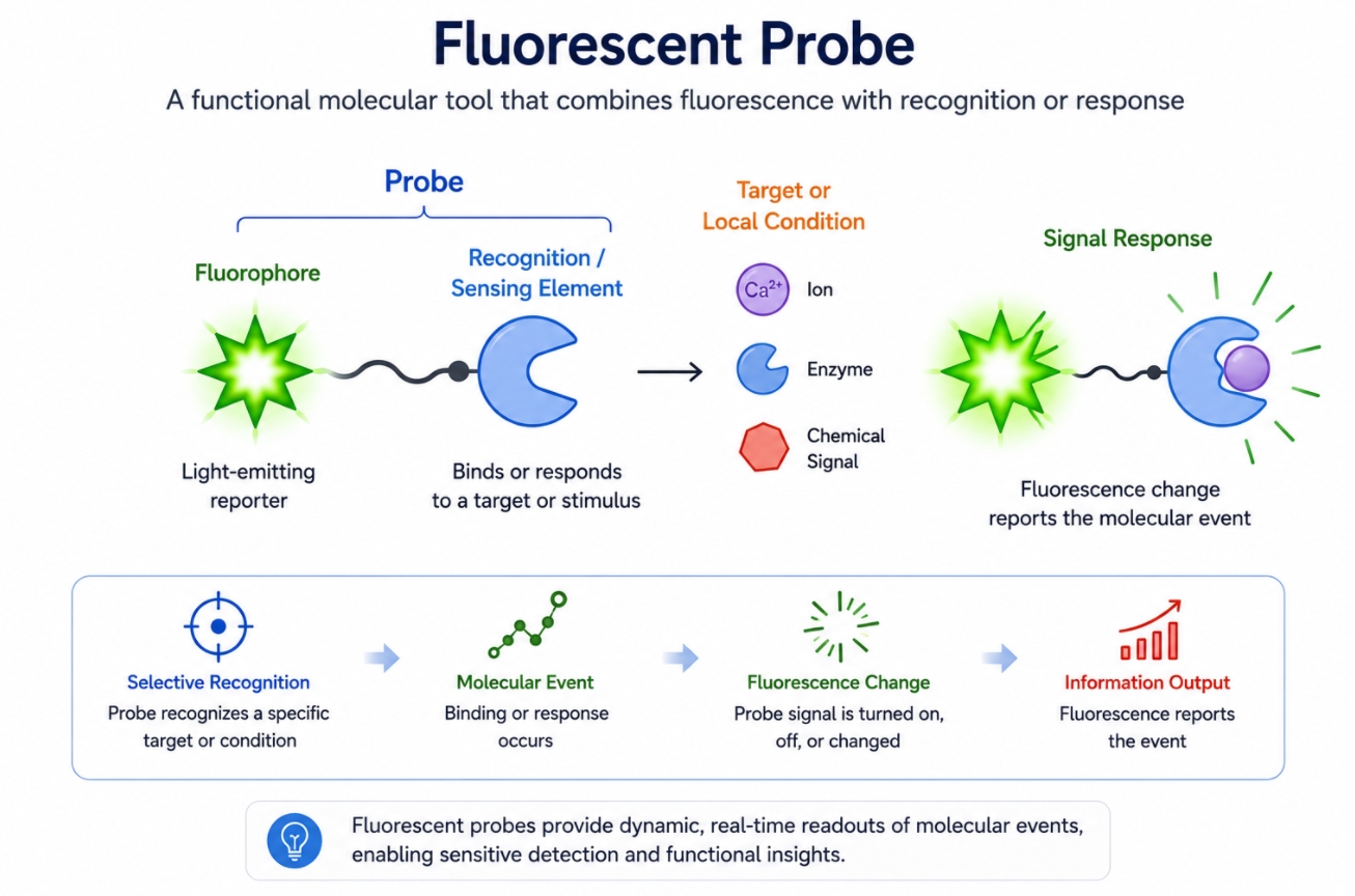 Fig. 3. Fluorescent probes combine fluorescence with recognition or response logic (BOC Sciences Authorized).
Probes Are More Than Carriers of Fluorescence
A fluorescent probe typically contributes functional meaning to the signal. The fluorescence is not only present; it is often linked to molecular recognition, local chemical conditions, or a change in probe state after interaction with the intended target or environment. This is what separates a probe from a simple dye that only supplies color or brightness.
- Recognition-Driven Signal: Many probes are designed to bind a target, recognize a molecular class, or respond to a defined biochemical feature. The useful signal therefore depends on the probe encountering the correct molecular context rather than simply being present in the sample.
- Responsive Readout: Probe fluorescence may increase, decrease, shift, or localize differently depending on pH, polarity, enzyme activity, reactive species, ions, or binding events. This makes the signal more information-rich, but also more dependent on assay conditions and careful interpretation.
- Functional Interpretation: The signal is often expected to carry mechanistic or conditional meaning, not just indicate that a fluorophore is present in the system. In practical terms, probe data are usually interpreted as evidence of a chemical or biological event rather than simple target occupancy.
- Different from a Simple Dye: A dye can become part of a probe, but a probe usually includes additional logic beyond fluorescence alone. That extra logic may come from a recognition motif, a triggerable scaffold, a responsive linker, or a built-in signal-modulation mechanism.
Structural and Functional Logic of Fluorescent Probe
From a design perspective, probes often contain more than one functional element. Their performance depends not only on fluorophore brightness, but also on how the sensing, targeting, or recognition elements are coupled to the fluorescent scaffold and how that structure behaves in the real assay environment. Probe design is therefore usually more architecture-sensitive than simple labeling.
- Multi-Component Architecture: A probe may include a fluorophore plus a targeting motif, recognition group, responsive trigger, cleavable linker, or quenched precursor design. Each part contributes to how the signal is generated and how selective the final readout becomes.
- Signal Coupled to Function: The fluorescence behavior is often intentionally linked to binding, conversion, activation, or environmental change. This means the signal must be evaluated not only for optical strength but also for whether it tracks the desired biochemical event faithfully.
- More Demanding Design Requirements: Good probe performance depends on balancing recognition selectivity, response behavior, fluorophore properties, and background control. A probe can fail even if its fluorophore is bright, simply because the recognition or activation logic does not behave cleanly in the real system.
- Workflow Sensitivity: Probe performance is often more sensitive to assay conditions than simple labels because the readout depends on both chemistry and local biological context. Buffer composition, pH, competing analytes, reaction timing, and compartmental environment can all influence how the probe behaves.
When Probes Are More Informative Than Simple Labels
Probes are usually the better choice when the experiment is trying to learn something about activity, condition, or local molecular state rather than simple location alone. In these workflows, the information value of the signal matters as much as the visibility of the signal. The probe is not just marking a target; it is helping define what kind of event or condition is being measured.
- Functional Imaging: Useful when the question involves enzyme activity, oxidative state, ion flux, membrane behavior, or local biochemical changes. In these cases, a simple label may show where something is, but a probe is better suited to show what is happening.
- Environmental Readouts: Appropriate when fluorescence should reflect pH, polarity, redox status, or compartment-specific chemistry. These conditions often require signal behavior that changes with the local environment rather than remaining constant after installation.
- Mechanistic Studies: Better suited than simple labels when the user needs fluorescence to report a process rather than merely mark a target. This is especially important in workflows where the same structure may appear similar morphologically but differ functionally.
- Higher Information Density: A probe can provide more biologically meaningful output than a passive fluorescent tag when the assay is designed to exploit that responsiveness. In many advanced workflows, the value of a probe lies in its ability to convert fluorescence into chemically or biologically interpretable information.
What Fluorescent Staining Usually Means?
Fluorescent staining usually refers to workflows in which fluorescence is used to visualize structures, compartments, or sample components by staining them rather than by building a highly defined target-conjugation system. In many staining workflows, the main goal is practical visualization: to reveal nuclei, membranes, organelles, DNA, RNA, lipid-rich structures, cell populations, or broad sample architecture. Staining can be highly useful and highly informative, but it usually emphasizes visualization outcome more than conjugation design. This makes it particularly attractive in morphology-oriented workflows and in applications where fast, interpretable structure-level fluorescence is more important than exact labeling-site control.
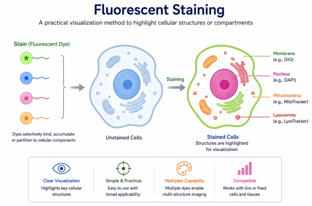 Fig. 4. Fluorescent staining highlights structures or compartments for practical visualization (BOC Sciences Authorized).
Staining Focuses on Visualizing Classes of Structures
The core logic of staining is often class-based rather than target-by-target. A stain is usually selected because it highlights a structure category or a sample feature broadly enough to support visualization, contrast generation, or structural interpretation. This makes staining especially useful when the experiment needs context, morphology, or rapid image readability rather than a highly engineered fluorescent association.
- Class-Level Visualization: Staining is often used for nuclei, membranes, organelles, nucleic acids, lipid-rich regions, or whole-cell populations rather than one uniquely defined molecular target. The signal is therefore interpreted at the level of a structural class rather than a single target identity.
- Strong Practical Utility: It is especially helpful when the goal is structural context, morphology, compartment contrast, or sample-level visualization. In many workflows, this kind of broad visual information is what makes later specific signals easier to interpret.
- Less Emphasis on Site Precision: The user is usually not asking where one exact labeling site exists, but whether the target class becomes visible clearly and reproducibly. This is why staining can be technically simpler while still being highly useful.
- Common in Imaging Workflows: Staining is widely used in microscopy, sample preparation, and comparative visualization tasks where rapid readout is important. It often provides the structural reference layer that allows more target-specific labeling or probe signals to be interpreted in context.
Staining Often Relies on Affinity or Partitioning
Unlike many target-directed labeling workflows, staining often depends on broad interaction logic rather than on covalent or engineered target association. This can make staining operationally simpler, but it also means the source of selectivity is different from that of labeling or probe-based recognition. The fluorescence becomes useful because the reagent distributes in a predictable way, not necessarily because it is attached to one defined molecular site.
- Affinity-Based Staining: Some stains bind preferentially to particular structural classes such as nucleic acids or membrane-associated components. The signal depends on preferential interaction rather than on a designed conjugation event.
- Partition-Driven Localization: Other stains accumulate because their physicochemical properties favor a certain compartment or environment. This partitioning logic is especially common in membrane-oriented or organelle-associated staining systems.
- Intercalation or Accumulation: Fluorescence may arise through insertion, distribution, or retention rather than through precisely defined attachment chemistry. This distinction is important because it changes how the signal should be interpreted and validated.
- Different from Controlled Conjugation: The signal can still be useful and reproducible, but it is usually not built on the same target-specific logic as a defined labeling workflow. Customers should therefore not expect the same type of molecular precision from a stain as from a target-directed fluorescent conjugate.
When Staining Is the More Practical Choice
Staining is often the most efficient option when the biological question is structural and the workflow benefits from a faster, more direct route to fluorescence. It is especially appropriate when the user needs contrast and visualization more than engineered fluorescent association. In these contexts, staining provides strong practical value without requiring the design burden of a custom target-labeling strategy.
- Morphology and Compartment Imaging: Useful when the goal is to visualize cellular architecture, organelle distribution, or structural contrast. In these workflows, fast and reliable image readability is often more important than exact target-level fluorescent attachment.
- Rapid Sample Assessment: Appropriate for workflows that require quick visualization of whether the sample has the expected structures or integrity. This can be particularly useful in imaging setup, QC-like checks, or early-stage workflow screening.
- Population-Level Visualization: Helpful when users need to distinguish cell groups or broad structural classes rather than individual molecular targets. Staining can provide a fast overview layer that complements more specific fluorescence strategies.
- Operational Simplicity: Often preferable when the workflow needs clean fluorescence with less conjugation design burden than a custom labeling route. For many customers, staining is the right starting point when a project needs visual utility first and molecular engineering second.
Key Differences Between Labeling, Probes, and Staining
Although fluorescent labeling, fluorescent probes, and fluorescent staining often overlap in real applications, they are not interchangeable concepts. The clearest way to distinguish them is to compare their design logic, specificity source, workflow structure, and the type of information they are usually meant to produce. The table below does not eliminate every overlap, but it provides a practical framework for deciding which term and which strategy are most appropriate in a real experimental context.
| Comparison Aspect | Fluorescent Labeling | Fluorescent Probes | Fluorescent Staining |
|---|---|---|---|
| Core Purpose | To introduce fluorescence into a defined target through a chosen labeling route. | To report, recognize, or respond to a target, state, or local condition using fluorescence. | To make a structure, compartment, or sample component visibly fluorescent for practical visualization. |
| Technical Logic | Target-directed installation of fluorescence. | Function-oriented molecular reporting or recognition. | Visualization-oriented staining or signal accumulation. |
| Specificity Source | Usually comes from conjugation chemistry, antibody recognition, genetic fusion, or controlled target installation. | Usually comes from molecular recognition, responsive chemistry, or environment-sensitive design. | Usually comes from affinity, partitioning, accumulation, or class-level structural preference. |
| Target Definition | Often a defined molecule, cell type, particle, or engineered construct. | Often a target plus a functional state, analyte, or microenvironment. | Often a structural class such as membranes, nuclei, nucleic acids, or organelle-rich regions. |
| Information Output | Often answers where the target is and whether it has been labeled successfully. | Often answers what the target is doing or what chemical state is present. | Often answers what structures are present and how they are distributed visually. |
| Typical Workflow Design | May involve conjugation, recognition-based detection, or staged fluorescent introduction. | Often built around a recognition or response mechanism plus fluorescence reporting. | Often emphasizes staining conditions, retention, contrast, and sample compatibility. |
| Typical Reagent Structure | Can range from simple fluorophore conjugates to antibody labels and engineered fluorescent constructs. | Often includes a fluorophore plus targeting, sensing, or responsive elements. | Often includes dyes or staining reagents chosen for affinity, localization, or partition behavior. |
| Degree of Structural Control | Often moderate to high, depending on labeling method. | Often moderate to high when probe design is mechanistically defined. | Often lower in positional control, but highly practical in structural visualization. |
| Speed and Convenience | Can vary widely depending on route complexity. | Can range from simple to complex depending on sensing architecture. | Often among the faster and more direct fluorescence workflows. |
| Best-Fit Applications | Conjugate development, target visualization, controlled fluorescent attachment, tracking. | Functional readouts, sensing, responsive imaging, mechanistic detection. | Cell and tissue visualization, morphology, compartment imaging, rapid structure highlighting. |
| Main Limitations | May require more chemistry control, validation, or target engineering. | May require more sophisticated design and interpretation than simple labels. | May provide less target-definition precision than controlled labeling routes. |
| Common Overlap Zones | Can overlap with probes or stains depending on reagent design and workflow context. | Can also function as labels or stains in some applications. | Can sometimes be described as labeling, especially in broad teaching or catalog language. |
Not sure whether your workflow needs labeling, probes, or staining?
We can help match the right fluorescence strategy to your target, readout, and experimental design.
Which One Should You Choose for Your Workflow?
The right choice depends less on terminology preference and more on what the experiment is actually trying to learn, how the signal needs to be generated, and how much control is required over reagent behavior and data interpretation. In practice, many users benefit from first deciding whether the project is primarily target-defined, function-defined, or structure-defined. That distinction usually determines whether labeling, probes, or staining should lead the workflow design. In more advanced systems, two or even all three may be combined, but they should still be chosen for different reasons rather than treated as interchangeable fluorescence tools.
Choose Labeling for Defined Targets and Controlled Conjugation
Fluorescent labeling is usually the strongest choice when the workflow begins with a defined target and the user wants fluorescence to remain explicitly associated with that target through a controlled route. This is especially important in protein conjugation, antibody labeling, nucleic acid derivatization, ligand tracking, engineered constructs, and custom assay development where the fluorescent signal must reflect a deliberate molecular installation event rather than general signal accumulation. From a technical perspective, labeling is advantageous when structural control, conjugation stoichiometry, fluorophore placement, and target retention all matter to downstream interpretation. Customers developing reusable reagents, target-tracking assays, or multi-step fluorescence workflows often benefit from a labeling strategy because it provides a stronger framework for reproducibility, validation, and integration with specific detection platforms. In other words, labeling is most appropriate when the question is not only "can I see fluorescence," but "can I control how fluorescence becomes associated with this target?"
Choose Probes for Functional or Responsive Readouts
Fluorescent probes are usually the better choice when the key question involves state, activity, local chemistry, or selective molecular recognition rather than simple target presence. If the project aims to monitor enzyme function, oxidative conditions, ion concentration, membrane state, pH, polarity, or another dynamic chemical feature, a probe-like design often provides much more useful information than a passive label. The technical strength of a probe is that the fluorescence is coupled to a mechanism, not merely to the presence of a fluorophore. From a customer perspective, this matters because probe-driven workflows often require a different kind of decision-making: not only which fluorophore is brightest, but which reagent architecture generates the most meaningful response under the real biological conditions of the assay. Probes are therefore especially valuable in functional imaging, mechanistic studies, sensing workflows, and any project where fluorescence is expected to report a process or condition rather than simply identify a predefined molecular target.
Choose Staining for Fast Structural Visualization
Fluorescent staining is often the most practical option when the main objective is rapid, interpretable visualization of structures, compartments, or broad sample features rather than tightly defined target engineering. This includes workflows focused on morphology, cell compartment contrast, nucleic acid visualization, membrane imaging, population-level structure identification, or basic sample assessment before deeper analysis. From a technical standpoint, staining is especially useful when speed, visual contrast, and workflow simplicity matter more than exact fluorescent attachment logic. For customers, staining is often the right choice when the project does not justify the development burden of a custom labeling strategy and does not require the functional reporting behavior of a probe. It is particularly effective in imaging workflows where structural context is essential, where broad classes of features need to be visualized efficiently, or where staining will be combined with more specific labels or probes to provide reference information in the same sample.
Combine Them When the Workflow Needs More Than One Layer
Many advanced fluorescence workflows are strongest when labeling, probes, and staining are combined deliberately rather than used in isolation. A user may employ fluorescent labeling to track a defined biomolecule, a probe to report local biochemical state, and a stain to reveal nuclei or membrane structure in the same experiment. In these situations, the categories do not collapse into one another; instead, they contribute different layers of information. From a technical perspective, combining them successfully requires careful channel planning, compatible fluorophore selection, and a clear understanding of what each signal is meant to represent. For customers designing multiplex or information-rich workflows, this layered strategy is often the best way to balance structural context, target-defined readout, and functional information in one integrated assay. The key is to assign each fluorescence component a clear role so that the final readout remains interpretable rather than becoming a visually complex but conceptually confused signal set.
How These Differences Affect Real Workflow Decisions?
From a customer perspective, the distinction between labeling, probes, and staining becomes most important when it changes what kind of reagent should be selected, how much control the workflow requires, and what kind of information the final signal is expected to support. In real projects, the wrong conceptual starting point often leads to the wrong development path: users may purchase a stain when the workflow actually requires a defined labeling route, or choose a probe-like reagent when the real need is fast structural contrast. These differences are not only semantic. They influence fluorophore selection, reagent architecture, assay setup, control design, data interpretation, and how easily the workflow can be optimized or reproduced later. The decision points below are often where the practical differences between these three fluorescence approaches become most meaningful.
- Defined Target vs Broad Structural Need: If the project starts with a known target that must carry fluorescence in a controlled way, fluorescent labeling is usually the better route. This is especially true when the user needs the signal to remain associated with a protein, antibody, nucleic acid, ligand, or engineered construct through a defined installation method. If the project instead begins with the need to visualize nuclei, membranes, compartments, lipid-rich regions, or cell populations more broadly, staining is often more practical because it emphasizes structural visibility rather than target-specific fluorescent attachment. The key technical difference is whether the workflow demands target-level control or class-level visualization.
- Static Visualization vs Functional Reporting: If the user only needs to know where something is, a label or stain may be sufficient depending on whether the target is defined or structural. If the user needs fluorescence to change meaningfully with chemical state, molecular activity, binding status, or local environment, a probe is usually the better technical choice. This distinction matters because functional readouts require more than fluorescent presence alone. They require a reagent whose signal is mechanistically linked to the event being studied. In practical terms, a static fluorescent image and a responsive fluorescent readout may look similar on screen, but they answer very different biological questions.
- Workflow Complexity and Development Burden: Labeling and probe-based workflows often require more design control, validation, and optimization than staining. A labeling workflow may require decisions about conjugation chemistry, fluorophore loading, purification, and target retention. A probe-based workflow may require even more careful evaluation because recognition logic, activation mechanism, background behavior, and assay conditions all affect whether the signal remains meaningful. Staining is often operationally simpler because it relies more on affinity, accumulation, or structural preference, but that simplicity usually comes with less target-definition control. Customers should therefore choose the more complex route only when the information gain justifies the added design and validation burden.
- Interpretability of the Final Signal: One of the most important practical questions is what the fluorescent signal is supposed to prove. A stain may show structure clearly but cannot always support strong target-level conclusions. A probe may report a condition, state, or response, but it may not provide strict molecular identity unless that logic is built into the design. A label may provide stronger target association, but by itself it usually does not report function or local chemistry unless combined with another responsive element. For customers, this means signal brightness alone is never the only criterion. The more important issue is whether the fluorescence output is technically aligned with the type of conclusion the workflow is expected to support.
- Need for Combined Strategies: In many real workflows, the best outcome comes from combining these concepts rather than choosing only one. A user may apply fluorescent labeling to track a defined biomolecule, use staining to provide structural context, and include a probe to report a relevant biochemical state in the same system. This layered approach can be highly informative, but only when each fluorescence component has a clearly defined purpose. Understanding the distinction between labeling, probes, and staining helps assign those roles correctly and reduces the risk of building an assay that is visually rich but technically ambiguous. From a workflow-design perspective, combination strategies are often most successful when channel planning, signal hierarchy, and interpretation logic are considered from the beginning rather than added reactively later.
How BOC Sciences Supports Fluorescent Labeling, Probe Selection, and Staining Workflows?
Fluorescence-based projects often require more than choosing a dye from a catalog. In practice, users may need to decide whether the workflow should be built around target-directed fluorescent labeling, a functional fluorescent probe, a structure-oriented staining strategy, or a combination of these. They may also need support in selecting suitable fluorophores, matching reactive groups to target chemistry, identifying probe architectures that fit the intended readout, or choosing staining reagents that provide clear contrast without creating unnecessary background. BOC Sciences supports these needs through both product supply and fluorescence-related service capabilities. Our goal is to help customers move from a general fluorescence idea to a more suitable combination of reagents, workflow logic, and development support for real research use.
Products for Fluorescent Labeling, Probes, and Staining
- Supply of fluorescent dyes, reactive fluorophore derivatives, probe-relevant building blocks, and staining-oriented reagents for a broad range of fluorescence workflows.
- Product categories relevant to target-directed fluorescent labeling, function-oriented probe development, and structure-focused staining applications.
- Reagent options for proteins, antibodies, peptides, nucleic acids, membranes, organelles, and other systems where fluorescence must serve different technical roles.
- Better product matching for users who need to distinguish whether the project calls for controlled labeling, responsive probe behavior, or practical staining contrast.
Fluorescent Labeling Service Support
- Support for selecting suitable fluorescent labeling routes according to target type, accessible chemistry, and required level of control over fluorophore installation.
- Assistance with matching fluorophore family, reactive group, and conjugation logic to proteins, antibodies, peptides, nucleic acids, and related target systems.
- Guidance on balancing signal strength, target compatibility, labeling density, and downstream assay requirements in custom or semi-custom labeling projects.
- Development-oriented support for fluorescent labeling workflows that must remain reproducible, technically interpretable, and adaptable to later optimization.
Probe and Staining Workflow Support
- Assistance with deciding whether a project is better served by a fluorescent probe strategy, a staining strategy, or a combined fluorescence workflow.
- Support for selecting fluorophore and reagent combinations that fit functional readouts, responsive fluorescence behavior, or structure-focused staining applications.
- Practical guidance for improving signal relevance, contrast quality, and workflow compatibility in probe-based or staining-based experiments.
- More suitable workflow planning for customers who need fluorescence to report state, reveal structure, or support both roles in the same system.
Custom Development and Integrated Fluorescence Solutions
- Support for custom fluorescence projects that require more than an off-the-shelf choice, including combined labeling, probe, and staining designs.
- Assistance with integrating reagent selection, fluorophore choice, and workflow logic into a more coherent fluorescence strategy.
- Development support for users building multi-layer workflows in which target labeling, functional reporting, and structural visualization must work together.
- Broader project support aimed at improving reagent fit, signal interpretability, and technical consistency across fluorescence-based research workflows.
Do You Need A Consultation?
BOC Sciences supports fluorescent labeling strategy design, probe selection, staining workflow planning, and custom fluorescent development for research applications.
Representative Reagents Relevant to Labeling, Probes, and Staining
| Catalog | Name | CAS | Inquiry |
|---|---|---|---|
| A14-0053 | 5,10,15-tris(phenyl)corrole | 246231-45-8 | Bulk Inquiry |
| A16-0050 | C6 NBD Galactosylceramide | 170212-26-7 | Bulk Inquiry |
| A16-0163 | 3,3'-Dipropyloxacarbocyanine iodide | 53213-79-9 | Bulk Inquiry |
| A16-0171 | Octadecyl Rhodamine B Chloride | 65603-19-2 | Bulk Inquiry |
| F04-0002 | FAM amine, 6-isomer | 1313393-44-0 | Bulk Inquiry |
| R01-0006 | BDP 581/591 NHS ester | 654651-21-5 | Bulk Inquiry |
| F08-0007 | Pyrene azide 1 | 2135330-58-2 | Bulk Inquiry |
| R01-0018 | Cyanine3.5 NHS ester | 2231670-85-0 | Bulk Inquiry |
| F04-0007 | Fluorescein-Maleimide | 2228857-33-6 | Bulk Inquiry |
| F04-0029 | 2',7'-Dichlorodihydrofluorescein | 106070-31-9 | Bulk Inquiry |
| R01-0042 | AF594 activated ester, 5-isomer | 1638544-48-5 | Bulk Inquiry |
| A03-0016 | Oregon Green 488 carboxylic acid | 195136-52-8 | Bulk Inquiry |
| F02-0003 | Cyanine3 carboxylic acid (chloride) | 1144107-76-5 | Bulk Inquiry |
| F03-0004 | Sulfo-Cyanine3 maleimide | 1656990-68-9 | Bulk Inquiry |
High-Performance Fluorescent Tools for Your Research
- Cyanine7 Near-infrared probes for optical imaging.
- Pyrene Dyes Environmental polarity sensing and membrane studies.
- Cyanine3 Standard green-orange fluorescent biomolecular labeling.
- Cyanine Versatile fluorophores for bioimaging applications.
- sulfo-Cyanine5 Hydrophilic red fluorescent bioconjugation.
- Cyanine3.5 Orange-red fluorescence for multiplex imaging.
- BODIPY Photostable dyes for lipid and cell imaging.
- sulfo-Cyanine3 Hydrophilic green-orange fluorescent conjugation.
Explore More Topics
Online Inquiry
