

How Fluorescent Proteins Overcome Live-Cell Protein Visualization Challenges?
Live-cell protein visualization technology is a fundamental basis of modern cell biology. It enables scientists to observe in real time the localization, dynamic changes, and functional states of proteins within cells, thus gaining deeper understanding of the molecular mechanisms in life processes. However, the complex environment of living cells often brings multiple challenges such as weak signals, high background noise, photobleaching, and phototoxicity, which limit the effectiveness of traditional imaging techniques. Fluorescent proteins (FPs), as genetically encoded endogenous labeling tools, play an important role in alleviating these bottleneck problems due to their diverse optical properties and high specificity.
What Is Protein Visualization?
Protein visualization refers to the use of various experimental techniques to present proteins within cells or organisms in intuitive image forms, thereby revealing their spatial distribution, dynamic changes, and functional states. Proteins, as functional executors in cells, participate in key biological processes such as signal transduction, cellular structure construction, and metabolic regulation. Accurate observation of protein expression, localization, and interactions is crucial for understanding cellular life activities and disease mechanisms.
 Fig. 1. Protein visualization in live-cell (BOC Sciences Authorized).
Definition and Importance in Modern Cell Biology
In modern cell biology, protein visualization technology not only allows scientists to observe cellular behavior from a macroscopic perspective but also reveals microscopic mechanisms of cellular functions at the molecular level. Through protein visualization, researchers can precisely track dynamic changes of proteins inside cells, identify abnormal protein aggregation or mislocalization, thereby promoting new drug development, disease diagnosis, and biotechnological applications.
Key Techniques for Visualizing Proteins in Living Cells
There are many protein visualization techniques, but dynamically observing proteins within living cells poses greater technical difficulties and challenges. The main technical methods include:
- Immunofluorescence: Uses antibodies to label proteins combined with fluorescent probes for imaging. However, cells generally need to be fixed, making this unsuitable for live-cell dynamic imaging.
- Fluorescent Protein Tagging: Through genetic engineering, target proteins are fused with fluorescent proteins, enabling real-time endogenous expression and observation. This has become the mainstream technology for live-cell protein visualization.
- Luciferase Reporter Systems: Monitor gene expression through bioluminescent enzyme reactions, suitable for overall functional analysis.
- Super-Resolution Microscopy: Breaks through optical resolution limits to achieve nanoscale protein localization, and combined with fluorescent proteins, further enhances live-cell imaging capabilities.
- Single-Molecule Tracking: Captures movement trajectories of individual protein molecules via highly sensitive detection.
Main Challenges in Live-Cell Protein Visualization
- Need for High-Specificity Labeling: There are numerous and complexly distributed proteins in living cells, making precise labeling of target proteins the foremost challenge in live-cell imaging. Traditional chemical dye labeling often lacks high specificity and involves complex procedures, making real-time tracking of dynamic proteins difficult.
- Biocompatibility and Cytotoxicity Issues: Imaging probes or labels must safely enter cells and stably express or exist without interfering with normal cellular functions. Many chemical dyes or exogenous labels may cause cytotoxicity, limiting their application in living cells.
- Limitations in Photostability and Signal Intensity: Live-cell imaging requires labels to maintain strong and stable fluorescence signals under prolonged excitation to track protein dynamics. Many traditional fluorescent dyes are prone to photobleaching, causing signal decay and affecting imaging quality.
- Conflict Between Spatial Resolution and Real-Time Imaging: Protein localization and movement within living cells usually occur at nano- to micrometer scales. Imaging technology must balance high spatial resolution with real-time dynamic imaging, posing high demands on probe performance.
Unique Advantages of Fluorescent Proteins
Fluorescent proteins are a class of protein labeling tools that can be directly expressed inside cells through genetic encoding. Since the discovery of green fluorescent protein (GFP), fluorescent proteins and their variants have rapidly become one of the most important imaging tools in life sciences, with main advantages including:
- Genetic Encoding for High-Specificity Localization: Fluorescent protein genes can be fused with target protein genes to form fusion proteins, ensuring that fluorescence signals strictly originate from the target protein. Through genetic engineering, researchers can achieve precise labeling and expression of specific proteins in living cells without complicated chemical modifications.
- Excellent Biocompatibility: Derived from natural proteins, fluorescent proteins have good biocompatibility. Cells can normally express them without affecting cellular functions, avoiding cytotoxicity potentially caused by exogenous chemical dyes.
- Diverse Optical Properties to Meet Multiple Imaging Needs: As research progresses, the fluorescent protein family has continuously expanded, covering a broad range of excitation and emission wavelengths from ultraviolet to far-red, with rich colors. Multicolor fluorescent proteins allow researchers to label and observe multiple proteins simultaneously, facilitating the study of complex cellular processes.
- Improved Photostability: Although some fluorescent proteins still face photobleaching issues, improved variants have greatly enhanced photostability, suitable for long-term, continuous live-cell imaging.
Support Applications of Fluorescent Proteins from BOC Sciences
| Solutions | Description |
|---|---|
| Cell Imaging | Visualize cellular structures and dynamics with high-performance fluorescent proteins for clear and accurate imaging. |
| In Vivo Imaging | Enable non-invasive, real-time tracking of biological processes in living organisms using our advanced fluorescent proteins. |
| Molecular Imaging | Facilitate precise molecular visualization and monitoring of interactions with bright and stable fluorescent proteins. |
| Protein Staining | Achieve specific and vivid protein labeling with fluorescent proteins for enhanced detection and analysis. |
| Immunofluorescence Staining | Enhance immunofluorescence assays with our fluorescent proteins for sensitive and specific biomarker detection. |
Applications of Fluorescent Proteins in Live-Cell Studies
With their genetically encoded nature and diverse optical properties, fluorescent proteins have become essential tools for dynamic observation of proteins in living cells. Their broad application fields cover protein localization, interactions, gene expression, cellular dynamics, and organelle studies.
Protein Localization and Tracking
By genetically fusing fluorescent proteins to target proteins, researchers can observe in real time the spatial distribution and migration trajectories of proteins within cells. For example, studying the dynamic distribution of membrane proteins on the plasma membrane, assembly and disassembly of cytoskeletal proteins, and localization changes of chromosome-associated proteins during the cell cycle. Such real-time tracking not only reveals spatial specificity of protein functions but also helps elucidate intracellular material transport and signal transduction mechanisms.
Studying Protein-Protein Interactions
Fluorescent proteins also play a key role in investigating protein-protein interactions. Using Förster resonance energy transfer (FRET) technology, researchers label interacting proteins with two different-colored fluorescent proteins. When two proteins come within a certain distance, energy transfers from the donor fluorescent protein to the acceptor, causing specific fluorescence changes, thereby demonstrating physical contact. This technique is highly sensitive and can monitor dynamic interactions in living cells in real time, helping to reveal mechanisms of signal transduction complex formation and dissociation, and assembly of protein complexes—key processes in life sciences.
Monitoring Gene Expression in Real Time
By placing the fluorescent protein gene under the control of specific gene promoters or regulatory sequences, fluorescent proteins act as reporter genes that directly reflect the transcriptional activity and expression patterns of target genes. Researchers can observe in real time the timing and spatial distribution of gene expression across different cell types, developmental stages, or environmental stimuli. For example, in developmental biology, fluorescent protein reporter systems dynamically track fluctuations of key developmental genes, clarifying cell fate determination mechanisms. This method offers high sensitivity, non-invasiveness, and quantifiability, making it an important tool in gene expression research.
Investigating Cellular Dynamics and Signaling
Fluorescent proteins have irreplaceable value in studying cellular dynamics. They can label key proteins involved in cell division, migration, and intracellular transport, enabling researchers to observe details of cellular behavior and internal molecular activities in real time. For instance, labeling spindle proteins allows analysis of chromosome separation mechanisms during mitosis; labeling actin or microtubules reveals dynamics underlying cell migration and morphological changes. Dynamic imaging with fluorescent proteins provides an intuitive, real-time perspective for deeper understanding of cell function and behavior.
Organelle Visualization and Functional Studies
Fluorescent proteins can be specifically targeted to particular organelles within cells—such as mitochondria, nuclei, and endoplasmic reticulum—helping scientists visually monitor organelle morphological changes and functional states. For example, targeting fluorescent proteins to mitochondria enables monitoring mitochondrial morphology dynamics, membrane potential changes, and their roles in apoptosis; labeling nuclei aids research into protein distribution within the nucleus and regulation of nuclear architecture. Such organelle-specific labeling not only advances organelle biology but also provides important research tools for related diseases such as mitochondrial disorders and nuclear abnormalities in tumor cells.
Overcoming Challenges in Live-Cell Protein Visualization
As a core tool for live-cell protein visualization, fluorescent proteins offer unique advantages by providing effective solutions to various bioimaging challenges.
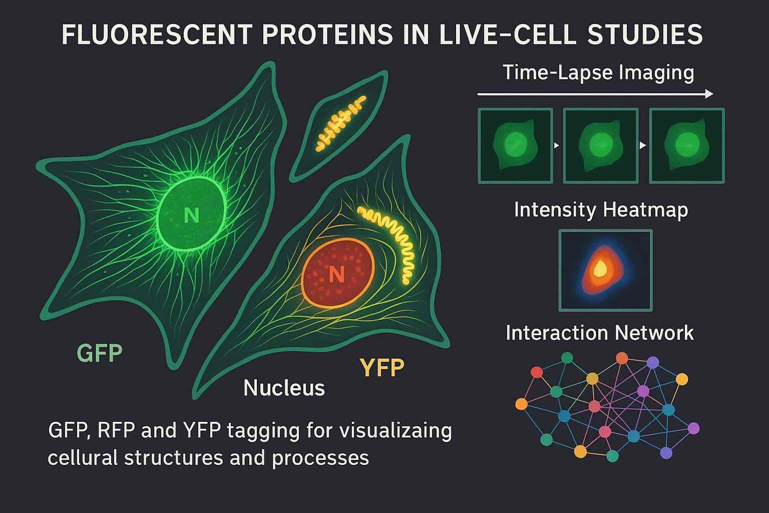 Fig. 2. Fluorescent proteins in live-cell studies (BOC Sciences Authorized).
Achieving Dynamic Localization and Real-Time Monitoring
Fluorescent proteins enable specific labeling of target proteins through genetic fusion, allowing real-time observation of protein dynamics inside living cells. Traditional dyes cannot achieve this function, while fluorescent proteins can be expressed directly, avoiding complicated procedures and cellular damage. For example, using GFP to label receptor proteins allows dynamic tracking of their distribution on the cell membrane and endocytosis processes, aiding studies of spatiotemporal regulation in signal transduction. Moreover, photoactivatable fluorescent proteins can selectively activate proteins in specific regions, combined with time-lapse imaging to finely resolve protein movement trajectories, significantly enhancing the capability for dynamic protein monitoring in live cells.
Multicolor Imaging Reveals Protein Interactions and Complex Networks
The fluorescent protein family includes multiple colors, enabling simultaneous labeling of various proteins within cells for multicolor colocalization imaging. By fusing different target proteins with blue, green, red, and other fluorescent proteins, their distribution and interactions can be observed simultaneously. For example, studying spatial and temporal dynamics of multiple proteins in signaling pathways reveals protein complex assembly and functional regulation. Multicolor fluorescent protein imaging greatly enriches the toolkit for investigating complex intracellular protein networks and provides intuitive means to decipher life processes.
Improved Photostability for Long-Term Imaging
Protein engineering has improved fluorescent proteins to have higher brightness and stronger resistance to photobleaching. Compared with traditional dyes, their photostability is significantly enhanced, suitable for prolonged continuous excitation during live-cell imaging. Improved variants such as EGFP, mCherry, and mScarlet ensure persistent fluorescence signals, supporting dynamic tracking of processes like the cell cycle, differentiation, and stress responses. High photostability enables researchers to obtain more complete information on protein dynamics, improving imaging data accuracy and reliability.
Enhanced Imaging Compatibility for Various Microscopy Techniques
Fluorescent proteins cover a broad spectral range, compatible with many modern microscopy platforms such as confocal, multiphoton, and super-resolution microscopes. Different colored fluorescent proteins suit multiple excitation light sources, enabling multichannel imaging. The development of near-infrared fluorescent proteins breaks through tissue penetration limits for deep in vivo imaging. Additionally, fluorescent proteins can be combined with optogenetic tools, integrating imaging and functional regulation capabilities, advancing research in cell signaling and neuroscience, thus providing strong support for diverse live-cell imaging applications.
Overcoming Cytotoxicity and Biocompatibility Barriers
As endogenously expressed proteins, fluorescent proteins exhibit good biocompatibility and do not interfere with normal cell functions. Compared with exogenous dyes, fluorescent proteins require no complex staining or fixation steps, avoiding cell damage and toxicity, making them suitable for long-term live-cell imaging. Cells express fluorescent proteins under natural physiological conditions, ensuring physiological relevance and data accuracy. This feature makes fluorescent proteins ideal tools for studying protein functions and dynamic processes, widely used in live-cell imaging.
Our Fluorescent Protein Solutions for Your Research
BOC Sciences is dedicated to providing comprehensive, high-quality fluorescent protein solutions for life science researchers, helping clients overcome technical bottlenecks in live-cell protein visualization and achieve precise, efficient scientific outcomes. Our extensive product range, customized services, and professional conjugation and labeling technologies meet complex experimental needs.
Wide Range of Fluorescent Protein Variants for Diverse Needs
- Classic green fluorescent protein (GFP), cyan fluorescent protein (CFP), yellow fluorescent protein (YFP), and various red fluorescent proteins.
- Near-infrared fluorescent proteins suitable for deep tissue imaging and in vivo dynamic observation.
- Multiple spectral and color options supporting multiplex labeling and multichannel colocalization experiments.
- High brightness and photostability fluorescent proteins suitable for long-term live-cell imaging.
- Fluorescent proteins compatible with various expression systems, including mammalian cells, yeast, and insect cells.
Custom Engineering and Optimization Services
- Custom fluorescent protein sequence design, optimizing excitation and emission spectra to avoid spectral overlap.
- Enhancing photostability and quantum yield to extend imaging duration and improve signal intensity.
- Designing fusion proteins tailored to specific experimental conditions, ensuring labeling specificity and biocompatibility.
- Optimizing expression vectors and tag positions to minimize interference with cell functions.
- Providing protein engineering services for novel fluorescent proteins to meet cutting-edge research demands.
Conjugation and Labeling Solutions for Advanced Applications
- Diverse fluorescent protein conjugation methods, including genetic fusion expression and covalent conjugation technologies.
- Efficient labeling systems such as biotin-avidin and enzyme-catalyzed conjugation to enhance signal detection sensitivity.
- Support for multiplex fluorescent labeling and multi-target imaging, enabling complex cellular network analysis.
- Integration of chemical modification techniques to enhance fluorescent protein stability and intracellular distribution control.
- Professional technical support to assist in designing labeling schemes and imaging strategies tailored to specific research needs.
Do You Need A Consultation?
BOC Sciences integrates cutting-edge fluorescence technologies to accelerate your research, driving next-generation solutions for drug discovery and diagnostics.
Cutting-Edge Fluorescent Tags for Proteins
| Cat. No. | Product Name | CAS No. | Inquiry |
|---|---|---|---|
| A01-0019 | Fluorescein-dT Phosphoramidite | N/A | Inquiry |
| A16-0036 | Calcein Blue | 54375-47-2 | Inquiry |
| A01-0008 | Thioflavin T | 2390-54-7 | Inquiry |
| A01-0005 | Rhodamine B | 81-88-9 | Inquiry |
| A01-0010 | Basic Yellow 2 | 2465-27-2 | Inquiry |
| A01-0018 | 6-Fluorescein Serinol Phosphoramidite | 1275574-87-2 | Inquiry |
| A01-0020 | Yakima Yellow® Phosphoramidite | 502485-39-4 | Inquiry |
| F04-0026 | FAM-xtra Phosphoramidite | 2304636-67-5 | Inquiry |
| F04-0027 | Fluorescein II CEP | 1027512-13-5 | Inquiry |
| A01-0013 | VIC phosphoramidite, 6-isomer | 1414265-81-8 | Inquiry |
High-Performance Fluorescent Tools for Your Research
- Nuclear Fluorescent Probes DNA-binding dyes for nucleus visualization.
- Metal Fluorescent Probes Selective sensors for intracellular metal ions.
- Cytoskeleton Fluorescent Probes Probes for actin and microtubule visualization.
- Nitric Oxide (NO) & Reactive Oxygen Species (ROS) Probes for oxidative stress and signaling detection.
- Apoptosis Fluorescent Probes Probes detecting programmed cell death events.
- pH Indicators Fluorescent sensors for intracellular pH monitoring.
- Calcium, Chloride and Other indicators Fluorescent indicators for intracellular ion flux monitoring.
- Endoplasmic Reticulum Fluorescent Probes ER-targeted dyes for organelle structure analysis.
Explore More Topics
Online Inquiry
