

Comprehensive Guide to Fluorescent Proteins and Their Uses in Research
Fluorescent proteins have their own optical characteristics and are often used as versatile fluorescent labeling tools in the life science research fields of cell imaging, protein tracking, and molecular detection. By using fluorescent proteins of different colors and spectral properties, complex multiplex labeling and dynamic monitoring can be achieved, greatly improving the sensitivity and accuracy of experiments. Structural, performance, and application studies of fluorescent proteins provide researchers with a reference to help them choose the right fluorescent protein, optimize experimental conditions, and promote the application of novel research.
What Are Fluorescent Proteins?
Fluorescent proteins are a class of natural proteins capable of absorbing light energy at specific wavelengths and emitting fluorescence at different wavelengths. The earliest discovered fluorescent protein is the green fluorescent protein (GFP), derived from the jellyfish Aequorea victoria. Due to their unique optical properties, fluorescent proteins have become important tools in molecular biology and cell biology, widely used in cell labeling, protein tracking, studies of molecular interactions, and bioimaging.
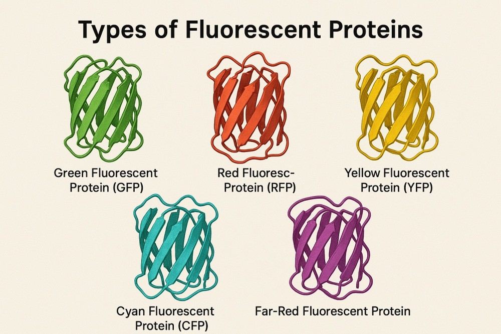 Fig. 1. Fluorescent proteins (BOC Sciences Authorized).
Fluorescent Protein Structure
The core structure of fluorescent proteins is a highly stable β-barrel conformation, composed of 11 β-strands forming a hollow barrel structure that encloses a special structure called the chromophore. The chromophore is usually formed spontaneously by chemical modification of several amino acid residues within the protein chain. This unique structure not only protects the chromophore from external environmental interference but also endows fluorescent proteins with high thermal and photostability, allowing stable luminescence even in complex cellular environments.
How Do Fluorescent Proteins Work?
Fluorescent proteins absorb excitation photons of specific wavelengths, causing electrons in the chromophore to transition to an excited state. When the electrons return to the ground state, they release emission photons of longer wavelengths. This process is called fluorescence emission. The difference between the excitation and emission wavelengths is known as the Stokes shift. Different fluorescent proteins have distinct chromophore structures, leading to variations in their excitation and emission spectra, resulting in diverse color options that enable multiplex labeling and multicolor imaging.
Why Are Fluorescent Proteins Important?
The importance of fluorescent proteins lies in their significant promotion of life science research. As endogenous fluorescent markers that can be expressed in living cells, fluorescent proteins avoid the cytotoxicity and permeability issues caused by traditional chemical dyes. They allow researchers to observe the localization, expression, and dynamic changes of proteins within cells in real time, dynamically, and non-destructively, greatly improving the accuracy of studies on cell functions and signaling pathways. Furthermore, the diversity of fluorescent protein colors and their genetic engineering potential provide strong support for advanced imaging techniques such as FRET and photoactivated localization microscopy, making them indispensable tools in modern molecular and cell biology.
Types of Fluorescent Proteins
Fluorescent proteins are diverse, covering a range of colors from ultraviolet to near-infrared wavelengths to meet different imaging and labeling needs in research. The optical properties of different fluorescent proteins are determined by their chromophore structures. After genetic engineering modifications, their colors and performances have been greatly enriched. The following are some common major types of fluorescent proteins:
Green Fluorescent Protein
Green fluorescent protein is the earliest discovered and widely applied fluorescent protein, originating from the jellyfish Aequorea victoria. Its excitation wavelength is about 488 nm, and emission wavelength is about 509 nm, showing bright green fluorescence. GFP has good thermal stability, photostability, and cell compatibility, making it a classic marker tool in cell and molecular biology research. Through directed evolution, multiple GFP mutants have greatly enriched its performance and application scope.
Cyan Fluorescent Protein
Cyan fluorescent protein is a GFP variant, with an emission wavelength around 475 nm, displaying blue-green fluorescence. CFP excitation is about 433 nm and is often used together with YFP or RFP for multicolor imaging and FRET (Förster Resonance Energy Transfer) experiments. Improvements in CFP's brightness and photostability have made it widely used in live-cell imaging and protein interaction studies.
Yellow Fluorescent Protein
Yellow fluorescent protein is another GFP variant, emitting light around 527 nm, presenting bright yellow fluorescence. YFP has a high quantum yield and good brightness, suitable for intracellular pH sensing, ion probes, and FRET techniques. Its longer emission wavelength reduces autofluorescence interference, helping to improve imaging contrast.
Red Fluorescent Protein
Red fluorescent proteins mainly originate from coral marine organisms, with emission wavelengths generally above 580 nm. Due to their longer emission wavelengths, RFPs have strong tissue penetration ability and low background autofluorescence, making them highly suitable for in vivo deep tissue imaging and multicolor colocalization studies. Common RFP family members include mCherry and DsRed. The photostability and brightness of RFPs have also significantly improved in recent years.
Blue Fluorescent Protein
Blue fluorescent protein emits light at about 445 nm and is an earlier fluorescent protein variant. Although its brightness and photostability are relatively low and it requires specific excitation wavelength and conditions, it still plays a unique role in some multicolor imaging experiments, especially when used in combination with other colored fluorescent proteins.
Other Fluorescent Proteins
Besides the above classic fluorescent proteins, researchers have developed various special-color and functional fluorescent proteins, such as orange fluorescent protein (OFP), violet fluorescent protein (VFP), and near-infrared fluorescent protein (iRFP). These proteins are often used in specialized applications like in vivo imaging, deep tissue penetration, and complex multiplex labeling experiments. Additionally, pH-sensitive, photoactivatable, and photoconvertible fluorescent proteins belong to special categories that respond to environmental stimuli for precise spatiotemporal labeling.
Support Applications of Fluorescent Proteins from BOC Sciences
| Solutions | Description |
|---|---|
| Molecular Imaging | Our fluorescent proteins enable precise visualization of molecular interactions for advanced imaging studies. |
| High-Throughput Screening | Enhance screening efficiency with fluorescent proteins as sensitive, reliable reporters in large-scale assays. |
| Drug Delivery | Track and monitor targeted drug delivery in real time using our versatile fluorescent proteins. |
| Cell Imaging | Illuminate cellular structures and dynamics with high-performance fluorescent proteins for clear cell imaging. |
| In Vivo Imaging | Our fluorescent proteins provide real-time, non-invasive imaging to monitor biological processes within living organisms. |
Fluorescent Protein Properties
The application effectiveness of fluorescent proteins highly depends on their physical and optical properties. Studying and selecting fluorescent proteins suitable for experiments requires a deep understanding of their brightness, color, and spectral characteristics to achieve optimal imaging results and data accuracy.
Fluorescent Protein Brightness
Brightness is an important indicator to evaluate fluorescent protein performance, defined as the product of its molar extinction coefficient and quantum yield. The molar extinction coefficient represents the protein's ability to absorb excitation light, while the quantum yield represents the efficiency of emitting fluorescence photons after absorbing photons. Higher brightness means stronger fluorescent signals, helping reduce excitation light exposure time and intensity, thereby lowering cellular phototoxicity and photobleaching risks. For example, the classic GFP has moderate brightness, while engineered superfolder GFP (sfGFP) shows significantly enhanced brightness. Different colored fluorescent proteins vary greatly in brightness, so both experimental needs and protein brightness must be considered when selecting.
Fluorescent Protein Colors
The color of fluorescent proteins refers to the emission wavelength range of their fluorescence, covering multiple bands from ultraviolet, blue, green, yellow, orange, red to near-infrared. The diversity of colors allows fluorescent proteins to realize multiplex labeling, support multichannel imaging, and complex intracellular process analysis. The colors are designed based on chromophore structural differences and regulated by gene mutations and protein engineering to tune the emission peak wavelength. For example, GFP emits green light (~509 nm), RFP emits red light (~600 nm), and combining different colored fluorescent proteins avoids signal overlap, ensuring experimental result accuracy.
Fluorescent Protein Spectra
The spectra of fluorescent proteins refer to their specific excitation and emission wavelengths of absorbed and emitted light. Each fluorescent protein has unique spectral characteristics determined by its chromophore structure, amino acid environment, and molecular conformation. Spectral characteristics are crucial in multicolor imaging, biosensing, and other fluorescence-based applications. Protein engineering techniques have expanded the spectral range of fluorescent proteins from ultraviolet (UV) to near-infrared (NIR) regions. This expansion allows multiple fluorescent proteins to be used simultaneously in complex experiments without obvious spectral overlap.
| Fluorescent Protein | Excitation Peak (nm) | Emission Peak (nm) | Color |
|---|---|---|---|
| GFP (Green) | ~488 | ~509 | Green |
| CFP (Cyan) | ~433 | ~475 | Cyan |
| YFP (Yellow) | ~514 | ~527 | Yellow |
| RFP (Red) | ~558 | ~583 | Red |
| mCherry | ~587 | ~610 | Red |
| mKate2 | ~588 | ~633 | Far-red |
| iRFP (Infrared) | ~690 | ~713 | Near-Infrared |
Fluorescent Protein Genes and Their Expression
The ability of fluorescent proteins to emit visible light within living organisms originates from their unique gene sequences and protein structures. The development of technologies for obtaining, modifying, and expressing these genes has made fluorescent proteins one of the most important molecular tools in modern life science research.
Overview of Fluorescent Protein Genes
Fluorescent protein genes originally come from certain marine organisms such as jellyfish (Aequorea victoria) and corals (genus Discosoma). Taking GFP as an example, its coding sequence was first successfully cloned and expressed in other organisms in the 1990s, ushering in a new era of fluorescent labeling technology. The original fluorescent protein genes have undergone extensive optimization to adapt to different host expression systems. Modern fluorescent protein families (such as mCherry, mNeonGreen, iRFP, etc.) have been developed on this basis, exhibiting superior optical performance and biocompatibility. These optimizations include:
- Codon optimization: Adjusting codon preferences in the gene to better suit expression in higher eukaryotic cells (e.g., mammalian cells).
- Removal of unstable sequences: Deleting regions that cause mRNA degradation or low translation efficiency.
- Mutations to enhance performance: Introducing amino acid mutations to improve brightness, photostability, pH tolerance, and resistance to photobleaching.
- Expansion of spectral range: Developing various fluorescent proteins with different colors through directed evolution and random mutation, such as CFP (cyan), YFP (yellow), RFP (red), and near-infrared fluorescent proteins.
Fluorescent Protein Expression in Biological Systems
Fluorescent protein genes are usually introduced into target biological systems through transfection, transgenic animals, or cell lines. The expressed fluorescent proteins can serve as fusion tags of endogenous proteins, allowing real-time observation of their localization and dynamic changes. The choice of expression system must consider the host cell type, protein folding efficiency, and luminescence requirements. Currently, fluorescent proteins are widely used across systems ranging from prokaryotes to higher eukaryotes:
- Bacteria and yeast: Studying gene expression regulation and metabolic pathways.
- Plants: Observing organelle localization and plant tissue development.
- Mammalian cells: Conducting cell imaging, molecular interaction detection, and signaling pathway research.
- Transgenic animals: Such as GFP mice and RFP zebrafish, used in developmental biology, neuroscience, and disease model studies.
Comparison and Selection of Fluorescent Proteins
With the continuous enrichment of fluorescent protein types, scientists face an important question in experimental design: how to choose the most suitable fluorescent protein type for specific applications. Comprehensive comparison of different fluorescent proteins and mastering selection principles and detection methods are key to ensuring experimental success.
Fluorescent Protein Comparison
Different fluorescent proteins exhibit significant differences in physicochemical and photophysical properties. Researchers typically compare these parameters side-by-side using spectral graphs and fluorescent protein performance tables to select the most suitable fluorescent protein according to experimental needs. Key parameters to focus on include:
- Spectral properties: Each fluorescent protein has unique excitation and emission wavelengths. For example, GFP has an excitation wavelength of 488 nm and emission at 509 nm; mCherry emits in the red range around 610 nm. In multicolor imaging experiments, selecting fluorescent proteins with high spectral separation effectively reduces spectral overlap and signal crosstalk.
- Brightness: Brightness is determined by the extinction coefficient and quantum yield together; the higher the brightness, the stronger the fluorescence signal visibility. High-brightness fluorescent proteins (such as mNeonGreen) are especially useful in low-expression systems or imaging small structures.
- Photostability: Photostability reflects the ability of fluorescent proteins to resist photobleaching under continuous illumination. Proteins with high photostability (such as mScarlet) are suitable for long time-lapse imaging.
- Maturation time: Different fluorescent proteins require varying times after expression to form their chromophore. Fast-maturing variants are particularly important for studying rapid biological processes.
- pH sensitivity: Some fluorescent proteins undergo fluorescence quenching under acidic or alkaline conditions. pH-stable fluorescent proteins maintain consistent fluorescence signals in different cellular environments, while pH-sensitive variants (such as pHluorin) can be used as pH sensors.
- Oligomerization state: Monomeric fluorescent proteins (such as mEGFP) avoid abnormal aggregation when fused to target proteins, ensuring accuracy in localization studies.
How to Choose the Appropriate Fluorescent Protein?
The choice of fluorescent protein should be matched to experimental goals and technical constraints. By aligning fluorescent protein characteristics with experimental requirements, researchers can achieve the best signal-to-noise ratio, reduce experimental artifacts, and improve imaging accuracy. Practical suggestions include:
- Application needs: For deep tissue imaging in animal tissues, choose far-red or near-infrared fluorescent proteins (such as mKate2 or iRFP) due to their stronger tissue penetration and lower autofluorescence. For super-resolution microscopy, prioritize fluorescent proteins with high photostability and minimal blinking.
- Multicolor imaging: To achieve simultaneous imaging of multiple targets, select fluorescent protein combinations with non-overlapping emission spectra (such as GFP, mCherry, and TagBFP) to minimize signal crosstalk and simplify image analysis.
- Cellular environment: Consider fluorescent protein performance under specific environments like hypoxia, oxidative stress, or pH changes. Some red fluorescent proteins exhibit higher photobleaching resistance in oxidative conditions.
- Expression system: Different expression systems (bacteria, yeast, mammalian cells) may affect fluorescent protein performance. Codon optimization and promoter strength also influence expression levels and fluorescence intensity.
What Are Fluorescent Proteins Used For?
The discovery of fluorescent proteins has transformed life science research, enabling scientists to observe, track, and analyze molecular processes in living organisms in unprecedented ways. With their endogenous luminescence and highly tunable optical properties, fluorescent proteins are widely applied in cell imaging, molecular detection, and drug screening, becoming irreplaceable tools in modern biotechnology.
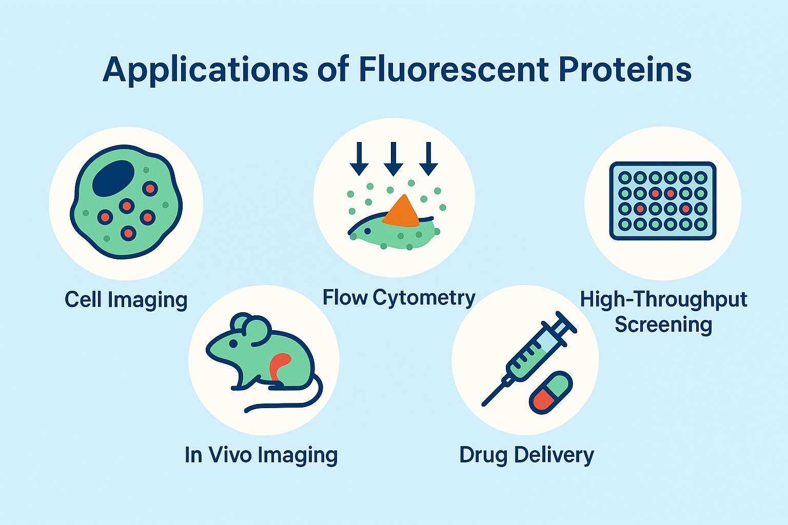 Fig. 2. Applications of fluorescent proteins (BOC Sciences Authorized).
Cell and Tissue Imaging
In cell and tissue imaging, the introduction of fluorescent proteins has made live imaging possible. By fusing fluorescent protein genes to specific target genes, researchers can observe the distribution and dynamic changes of target proteins within cells in real time. For example, GFP and its derivatives are widely used to reveal cytoskeletal structures, organelle morphology, and cell-cell interactions. Red and near-infrared fluorescent proteins, due to their longer emission wavelengths, are more suitable for deep tissue imaging and are used in animal models to study complex processes such as neural networks, tumor microenvironments, and organ development.
Protein Tracking and Localization Studies
Fluorescent proteins also play a key role in protein tracking and localization studies. By constructing fluorescent fusion proteins, researchers can monitor the spatial and temporal distribution of specific proteins during different developmental stages or external stimuli. For example, in studying cell signaling pathways, fluorescently labeled signaling molecules reveal their activation, transport, and degradation processes. This dynamic tracking ability provides strong support for understanding protein functions, interaction networks, and cellular behaviors.
Biosensors and Molecular Probes
Fluorescent proteins are central in the development of biosensors and molecular probes. Sensors based on Förster Resonance Energy Transfer (FRET) can detect ion concentrations, pH changes, or levels of small molecule metabolites, providing precise tools for intracellular microenvironment monitoring. For example, pH-sensitive fluorescent proteins (such as pHluorin) track acid-base changes in endocytic pathways, while calcium-sensitive fluorescent probes (such as the GCaMP series) are widely used in neuroscience for monitoring neuronal activity.
High-Throughput Screening in Drug Discovery
In the field of high-throughput screening for drug discovery, fluorescent proteins also demonstrate great application potential. By using fluorescent signals as experimental endpoints, researchers can rapidly evaluate drug effects on target pathways or proteins. For instance, expressing specific fluorescent reporter genes in cell lines allows signal intensity changes after drug treatment to reflect regulatory effects. This approach not only improves screening efficiency but also reduces costs and has become an important technology in modern pharmaceutical companies and research institutions.
Advanced Fluorescent Proteins for Specialized Applications
With the rapid development of bioimaging technologies and molecular tools, traditional fluorescent proteins no longer meet the demands of certain advanced experiments for spatiotemporal resolution, environmental responsiveness, and multicolor imaging. To address this, researchers have developed various advanced fluorescent proteins with unique functional properties, enabling them to play key roles in complex biological research and emerging applications.
pH-Sensitive Fluorescent Proteins
pH-sensitive fluorescent proteins are tools designed to respond to changes in intracellular acid-base environments. Many important cellular processes, such as endocytosis, exocytosis, and autophagy, involve significant pH fluctuations. pH-sensitive fluorescent proteins (such as pHluorin and pHRed) emit different fluorescence intensities at various pH values, allowing researchers to monitor in real time the acidification processes of organelles like lysosomes and endosomes. These fluorescent proteins are widely used in studies of viral invasion pathways, drug release mechanisms, and cellular metabolic states.
Photoactivatable Fluorescent Proteins
Another important class of tools is photoactivatable fluorescent proteins (PAFPs). They are initially non-fluorescent or weakly fluorescent but become activated into a high-intensity fluorescent state upon irradiation with specific wavelengths of light. PAFPs (such as PA-GFP and PA-mCherry) enable precise temporal and spatial labeling in live samples, greatly advancing studies of cell motility, synaptic plasticity, and developmental processes. Through light-controlled activation, scientists can track the dynamic behavior of individual cells or molecular populations without disturbing unlabeled regions.
Photoconvertible Fluorescent Proteins
Photoconvertible fluorescent proteins (PCFPs) undergo changes in emission color upon light exposure. For example, Kaede and Dendra2 can convert from green to red, while EosFP changes from green to orange-red. This characteristic makes them ideal tools for cell lineage tracing and time-resolved imaging. Researchers can "mark" a group of cells at a specific time point and then track their migration, differentiation, or interactions through changes in emitted light color. This precise spatiotemporal control is crucial for revealing complex biological phenomena such as developmental biology, tumor metastasis, and neural network remodeling.
Fluorescent Protein Products and Services
With years of R&D experience and a robust technical platform, BOC Sciences is dedicated to providing high-quality, diverse fluorescent protein products and comprehensive custom services to global research customers. We possess advanced gene synthesis and protein expression systems, combined with a professional protein engineering team and strict quality control processes, ensuring that every batch of fluorescent protein products exhibits excellent optical performance and biological stability.
Wide Range of Fluorescent Protein Variants Supplied
- A rich series of fluorescent proteins covering multiple colors and spectra, including classic GFP, CFP, YFP, RFP, BFP, and more.
- Products with different excitation and emission wavelengths to support multichannel and multicolor imaging experiments.
- High purity and high stability products to guarantee experimental reproducibility and reliability.
- Multiple packaging specifications to meet needs from small-scale research to large-scale production.
- Stable inventory and flexible delivery schedules to ensure project timelines are unaffected.
Custom Synthesis and Engineering Services
- Design and synthesis of fluorescent proteins according to customer requirements, including mutants and fusion proteins.
- Protein sequence optimization services to improve expression efficiency and protein stability.
- Engineering modifications to enhance optical performance, such as photostability and emission intensity.
- Custom tag and functional domain fusions to meet special research needs.
- Support for selecting protein expression systems and process development to facilitate efficient production.
Conjugation and Labeling Services for Fluorescent Proteins
- Various conjugation strategies, including covalent coupling and non-covalent binding techniques.
- Combination with diverse labeling molecules such as antibodies, small molecule probes, peptides, etc.
- Optimization of conjugation conditions to maintain protein activity and fluorescence performance.
- Custom complexes such as fluorescent protein–nanoparticle and fluorescent protein–antibody conjugates.
- Strict quality control to ensure the purity and functional consistency of conjugation products.
Do You Need A Consultation?
BOC Sciences integrates cutting-edge fluorescence technologies to accelerate your research, driving next-generation solutions for drug discovery and diagnostics.
Cutting-Edge Fluorescent Tags for Proteins
| Cat. No. | Product Name | CAS No. | Inquiry |
|---|---|---|---|
| A01-0019 | Fluorescein-dT Phosphoramidite | N/A | Inquiry |
| A16-0036 | Calcein Blue | 54375-47-2 | Inquiry |
| A01-0008 | Thioflavin T | 2390-54-7 | Inquiry |
| A01-0005 | Rhodamine B | 81-88-9 | Inquiry |
| A01-0010 | Basic Yellow 2 | 2465-27-2 | Inquiry |
| A01-0018 | 6-Fluorescein Serinol Phosphoramidite | 1275574-87-2 | Inquiry |
| A01-0020 | Yakima Yellow® Phosphoramidite | 502485-39-4 | Inquiry |
| A01-0006 | Proflavine Hemisulfate | 1811-28-5 | Inquiry |
| R01-0472 | Atto 425-NHS ester | 892156-28-4 | Inquiry |
High-Performance Fluorescent Tools for Your Research
- Cell Proliferation Tracer Fluorescent Probes Long-term tracking of cell division processes.
- Nerve Terminal Probes Fluorescent tracers for synaptic activity analysis.
- Endoplasmic Reticulum Fluorescent Probes ER-targeted dyes for organelle structure analysis.
- Ion Fluorescent Probes Indicators for real-time ion concentration imaging.
- Lysosomal Fluorescent Probes Acidic organelle markers for lysosome tracking.
- pH Indicators Fluorescent sensors for intracellular pH monitoring.
- Cytoskeleton Fluorescent Probes Probes for actin and microtubule visualization.
- Apoptosis Fluorescent Probes Probes detecting programmed cell death events.
Explore More Topics
Online Inquiry
