

How Fluorescent Nanoparticles are Transforming Bioimaging in Research?
As life sciences and medical research continue to advance, bioimaging technologies play a vital role in revealing cellular functions, molecular mechanisms, and disease diagnosis. However, traditional imaging methods often face numerous challenges due to insufficient sensitivity, limited resolution, and lack of real-time monitoring capabilities. Fluorescent nanoparticles are tiny particles ranging from 1 to 100 nanometers in size that can emit stable and intense fluorescent signals. Their emission wavelengths can be tuned by adjusting materials and sizes, and they possess high photostability and excellent biocompatibility. With these unique properties, fluorescent nanoparticles provide higher sensitivity, superior spatial resolution, and real-time dynamic monitoring capabilities for bioimaging, becoming an important tool for overcoming the limitations of conventional technologies.
What Are Fluorescent Nanoparticles?
Fluorescent nanoparticles are tiny particles typically in the size range of 1 to 100 nanometers that can emit visible or near-infrared fluorescence. They generally consist of a luminescent core and a protective shell, with the core containing luminescent substances such as organic dyes, quantum dots, rare earth elements, or metallic nanoparticles. Through specialized design, these nanoparticles exhibit high brightness, excellent photostability, and tunable emission wavelengths, making them widely used in biological labeling, imaging, and diagnostic applications.
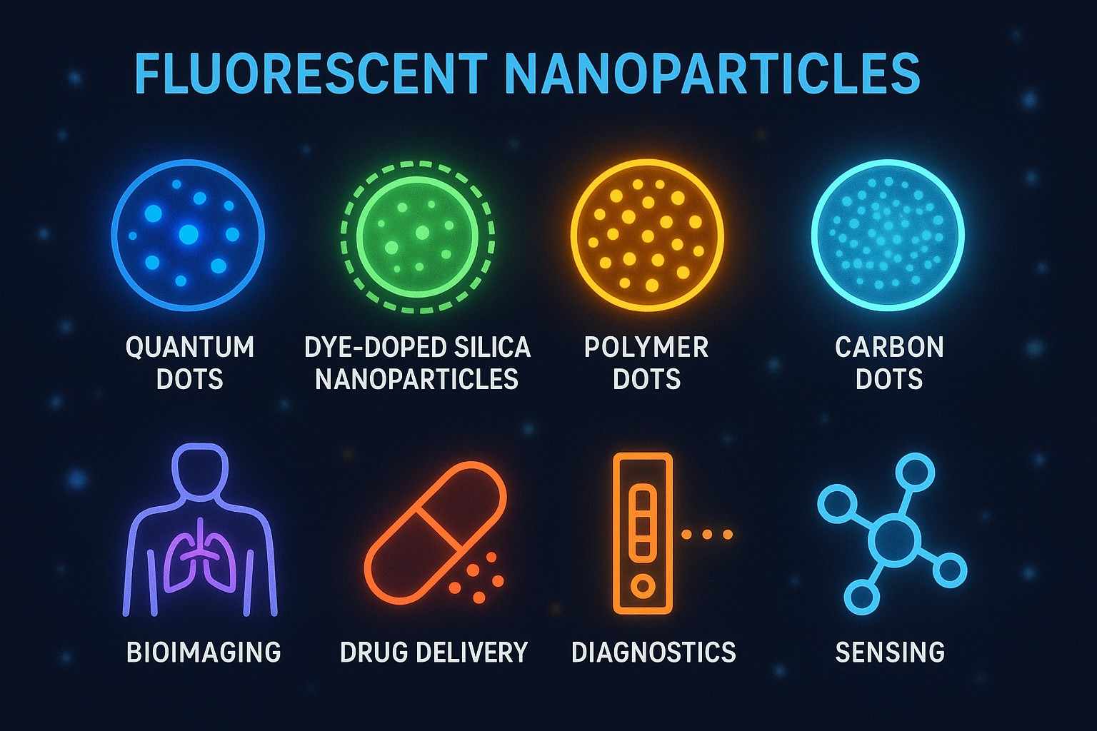 Fig. 1. Fluorescent nanomaterials and their applications (BOC Sciences Authorized).
Types of Fluorescent Nanoparticles
Fluorescent nanoparticles can be classified into various types based on their composition and luminescence mechanisms:
- Quantum Dots (QDs): Made of semiconductor materials, they feature broad excitation and narrow emission spectra, high fluorescence intensity, and good photostability, suitable for multicolor imaging.
- Rare Earth-Doped Nanoparticles: Utilizing the unique luminescence mechanisms of rare earth elements, they offer long lifetimes and resistance to photobleaching, ideal for long-term observation.
- Organic Fluorescent Nanoparticles: Typically lipid or polymer nanoparticles encapsulating organic dyes, with excellent biocompatibility.
- Metal Nanoparticles (e.g., gold nanoparticles): Enhance signals through surface-enhanced Raman scattering (SERS) effects, suitable for highly sensitive detection.
Why Use Fluorescent Nanoparticles for Bioimaging?
Compared to traditional fluorescent dyes and protein labels, fluorescent nanoparticles offer significant advantages, making them a key component of modern bioimaging technologies:
- High Brightness and Photostability: Fluorescent nanoparticles emit much stronger fluorescence than traditional organic dyes and are less prone to photobleaching, suitable for long-term tracking.
- Tunable Emission Wavelengths: By adjusting their material composition and size, they can cover a broad range from ultraviolet to near-infrared, enabling multicolor imaging.
- Multifunctional Surface Modification: Their surfaces can be easily conjugated with antibodies, ligands, or drugs for targeted recognition and functional applications.
- Good Biocompatibility and Low Toxicity: Modern synthesis techniques have significantly improved their biosafety, meeting the requirements for in vivo and in vitro applications.
Challenges in Biological Imaging Research
As a critical tool in modern life science research and medical diagnostics, bioimaging technologies have greatly advanced our understanding of cellular structures, molecular interactions, and disease mechanisms. However, despite technological progress, traditional bioimaging methods still face multiple challenges in practical applications, limiting their effectiveness and scientific value in complex biological systems.
Low Sensitivity in Conventional Imaging Techniques
Traditional fluorescent dyes and probes often suffer from weak signals and severe photobleaching when labeling and detecting biological samples. Many key molecules and cells involved in biological processes exist in very low quantities, and if the imaging system lacks sufficient sensitivity, it becomes extremely difficult to accurately capture signals from these low-abundance targets. In addition, background noise interference further reduces detection sensitivity. Low sensitivity not only affects image quality but also limits the discovery of early pathological changes and microscopic biological events, making it hard to deeply investigate many potential biomarkers and molecular mechanisms.
Poor Resolution in Complex Biological Samples
Biological samples, especially living tissues, are highly complex, containing multiple cell layers, abundant extracellular matrix, and inherent autofluorescence. These factors cause light scattering and signal attenuation, significantly reducing spatial resolution. Traditional optical microscopes are constrained by the diffraction limit of light, making it challenging to clearly resolve fine structures and molecular complexes inside cells. Particularly in deep tissue imaging, achieving both high resolution and sufficient imaging depth is difficult, affecting precise analysis of cellular functions and pathological changes.
Lack of Real-Time Monitoring
Many traditional bioimaging technologies are primarily based on static imaging of fixed samples and cannot continuously observe dynamic processes in live cells or tissues. This lack of real-time monitoring prevents researchers from capturing critical moments in dynamic biological processes such as cell migration, molecular interactions, and drug release, which impacts the understanding of biological mechanisms and the development of precise intervention strategies. Additionally, issues such as photobleaching and cell damage during prolonged imaging limit the feasibility of real-time dynamic imaging.
How Fluorescent Nanoparticles Solve Imaging Challenges?
Facing challenges such as low sensitivity, limited resolution, and lack of real-time monitoring in traditional bioimaging technologies, fluorescent nanoparticles provide powerful solutions due to their unique physicochemical properties and biological functional advantages. They not only significantly improve imaging performance but also advance in-depth research and precise diagnostics in complex biological systems.
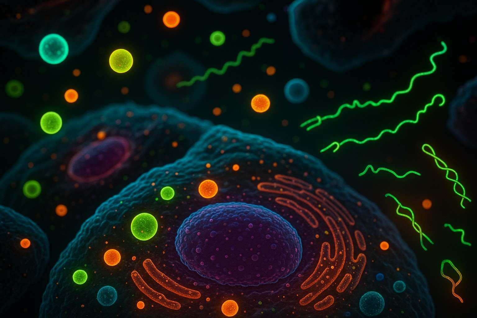 Fig. 2. Fluorescent nanoparticles in biological imaging (BOC Sciences Authorized).
Enhanced Sensitivity and Detection
Fluorescent nanoparticles such as quantum dots and rare earth-doped nanoparticles have extremely high quantum efficiency and strong luminescence signals, far surpassing traditional organic dyes. This allows them to produce detectable strong light signals even at very low concentrations, greatly enhancing the sensitivity of bioimaging. At the same time, their excellent photostability prevents photobleaching and signal attenuation, ensuring continuity and reliability during long-term imaging. Furthermore, the multicolor emission characteristics of nanoparticles enable simultaneous labeling of multiple molecules within the same sample, allowing multiplex detection and synchronized observation of complex biological processes. The combination of high sensitivity and multicolor imaging greatly expands their application potential in biological labeling and early disease diagnosis.
Superior Resolution for Cellular Imaging
The nanoscale size of fluorescent nanoparticles enables them to easily penetrate cell membranes and reach specific intracellular regions, precisely locating organelles, protein complexes, and other subcellular structures. Their high-brightness signals help overcome scattering and autofluorescence background in biological tissues, resulting in clear imaging outcomes. Coupled with advanced microscopy techniques (such as confocal microscopy and super-resolution microscopy), fluorescent nanoparticles can achieve spatial resolutions far beyond the optical diffraction limit, revealing detailed insights into intracellular microstructures and molecular interactions. This provides critical perspectives and data support for studying cellular functions, signal transduction, and pathological changes.
Real-Time Imaging Capabilities
Due to their photostability and low toxicity, fluorescent nanoparticles are particularly suitable for live-cell and in vivo imaging, supporting prolonged, continuous dynamic observation. They enable real-time monitoring of cell migration, molecular interactions, drug release, and pathological changes, capturing key moments that are difficult to observe with traditional techniques. The real-time imaging capability not only enhances understanding of the spatiotemporal dynamics of biological processes but also provides valuable information for drug development, disease diagnosis, and therapy monitoring. The integration of nanoparticles with highly sensitive imaging instruments has enabled a new mode of bioimaging characterized by high throughput, noninvasiveness, and high precision.
Support Applications of Fluorescent Nanoparticles from BOC Sciences
| Solutions | Description |
|---|---|
| Cell Imaging | Supporting precise cell visualization with custom-developed fluorescent nanoparticles for enhanced imaging quality. |
| In Vivo Imaging | Enabling deep tissue and real-time live imaging through tailored fluorescent nanoparticle solutions. |
| Molecular Diagnostics | Providing bright, specific fluorescent nanoparticles to improve sensitivity and accuracy in molecular diagnostics. |
| Fluorescence Microscopy | Delivering high-performance fluorescent nanoparticles optimized for superior microscopy imaging results. |
| Drug Delivery | Enhancing targeted drug delivery with custom fluorescent nanoparticles for precise tracking and controlled release. |
| Fluorescence Immunoassay | Supporting sensitive and specific immunoassays through tailored fluorescent nanoparticles for improved detection. |
Applications of Fluorescent Nanoparticles in Biological Imaging
With their excellent optical properties and biocompatibility, fluorescent nanoparticles have become an important tool in the field of biological imaging. They show great application potential in areas such as cell imaging, molecular detection, and in vivo imaging, driving progress in both basic research and clinical diagnostics.
Cell Imaging and Tracking
Fluorescent nanoparticles can achieve targeted labeling of specific cell types or organelles through surface modification with specific antibodies, peptides, or ligands. The high brightness and photostability of the nanoparticles enable researchers to track cell behaviors and movement trajectories over extended periods in complex biological environments. This technology is widely applied in fields such as tumor cell migration, stem cell differentiation, and immune cell dynamic monitoring. Through real-time imaging, researchers can observe cell proliferation, migration, and interactions, gaining deeper insights into disease development processes and therapeutic responses.
Molecular Detection and Diagnostics
Fluorescent nanoparticles play an important role in molecular-level detection and diagnostics. Utilizing their multicolor emission and high signal-to-noise ratio, multiple target molecules such as DNA, RNA, and proteins can be simultaneously detected, greatly enhancing detection sensitivity and specificity. As signal amplifiers, nanoparticles can be used in biosensors and immunoassays to support early disease diagnosis and precision medicine. For example, immunofluorescence techniques combined with fluorescent nanoparticles can quickly identify pathogens and tumor markers, providing critical information for clinical decision-making.
In Vivo Imaging
Fluorescent nanoparticles are particularly suitable for near-infrared emission, enabling noninvasive imaging of deep tissues. Their excellent light penetration ability and low background interference make it possible to locate tumors, track drug distribution, and perform tissue functional imaging in vivo. In live animal models, fluorescent nanoparticles can monitor disease progression and treatment effects in real time, supporting surgical navigation and the development of personalized treatment plans. By adjusting the surface functionalities of nanoparticles, targeted delivery and combined diagnosis and therapy can also be achieved, further expanding their clinical application prospects.
Our Fluorescent Nanoparticles for Bioimaging Services
As a leading provider of fluorescent reagents and custom services, BOC Sciences focuses on offering high-quality, high-performance fluorescent nanoparticles and related solutions to help advance bioimaging research to new heights. With advanced synthesis platforms and a professional technical team, we are committed to meeting customers' diverse research needs.
Custom Synthesis of Fluorescent Nanoparticles
- Precise control of nanoparticle size (1–100 nm range) to meet different imaging requirements;
- Multiple material options: quantum dots, rare earth-doped particles, organic fluorescent nanoparticles, etc.;
- Adjustable emission wavelengths covering ultraviolet to near-infrared, supporting multicolor imaging;
- High photostability and quantum efficiency to ensure imaging brightness and durability.
Surface Functionalization and Modification
- Diverse surface modification techniques: antibody conjugation, ligand modification, PEG coating, etc.;
- Enhanced biocompatibility and in vivo stability of nanoparticles;
- Optimization of nanoparticle dispersibility to prevent aggregation and improve experimental reproducibility;
- Design of functional groups according to customer requirements, supporting drug delivery and sensor development.
Real-Time Monitoring and Imaging Solutions
- Providing technical support for real-time dynamic monitoring in live-cell and in vivo imaging;
- Assisting in building high-sensitivity imaging platforms to capture cell migration and molecular interactions;
- Supporting continuous tracking and analysis of drug release processes;
- Offering imaging solution design, experimental process optimization, and data analysis services.
Do You Need A Consultation?
BOC Sciences integrates cutting-edge fluorescence technologies to accelerate your research, driving next-generation solutions for drug discovery and diagnostics.
Transform Your Studies with Cutting-Edge Fluorescent Products
| Cat. No. | Product Name | CAS No. | Inquiry |
|---|---|---|---|
| F01-0064 | meso-CH2Br-BODIPY | 216434-81-0 | Inquiry |
| A17-0186 | Perylene Orange | 82953-57-9 | Inquiry |
| A15-0005 | 5(6)-Carboxyfluorescein | 72088-94-9 | Inquiry |
| F04-0033 | 5-Aminofluorescein | 3326-34-9 | Inquiry |
| F04-0034 | 5-Carboxyfluorescein diacetate | 79955-27-4 | Inquiry |
| F04-0055 | Dexamethasone Fluorescein | 216854-76-1 | Inquiry |
| F02-0026 | Cy5-NHS ester | 146368-14-1 | Inquiry |
| F05-0031 | 6-Carboxy-X-rhodamine | 194785-18-7 | Inquiry |
| R02-0022 | Cyanine5 alkyne | 1223357-57-0 | Inquiry |
| F02-0007 | Cyanine5 amine | 1807529-70-9 | Inquiry |
High-Performance Fluorescent Tools for Your Research
- sulfo-Cyanine3.5 Water-soluble orange-red fluorescent labeling.
- BODIPY Photostable dyes for lipid and cell imaging.
- Cyanine3.5 Orange-red fluorescence for multiplex imaging.
- Cyanine7.5 Extended NIR imaging for in vivo studies.
- Cyanine Versatile fluorophores for bioimaging applications.
- Fluorescent Dyes General-purpose labeling for bioanalytical detection.
- Alexa Fluor Bright, photostable dyes for fluorescence imaging.
- sulfo-Cyanine5.5 Water-soluble far-red imaging probe.
Explore More Topics
Online Inquiry
