
Cell and Organelle Stains
- Mitochondrial Fluorescent Probes
- Lysosomal Fluorescent Probes
- Golgi Fluorescent Probes
- Endoplasmic Reticulum Fluorescent Probes
- Cell membrane Fluorescent Probes
- Nuclear Fluorescent Probes
- Cytoskeleton Fluorescent Probes
- Cell Proliferation Tracer Fluorescent Probes
- Apoptosis Fluorescent Probes
- Lipid Fluorescent Probes
- Nerve Terminal Probes
- Other Cell Fluorescent Probes
Customized Fluorescent Reagents
One-stop Solution for Your Research
BOC Sciences offers a one-stop solution for fluorescent reagents, providing custom synthesis, modification, and large-scale production services. Our comprehensive portfolio includes high-purity fluorescent dyes, probes, and labeling reagents for research and industrial applications, ensuring superior performance and reliability.
Explore More
Background
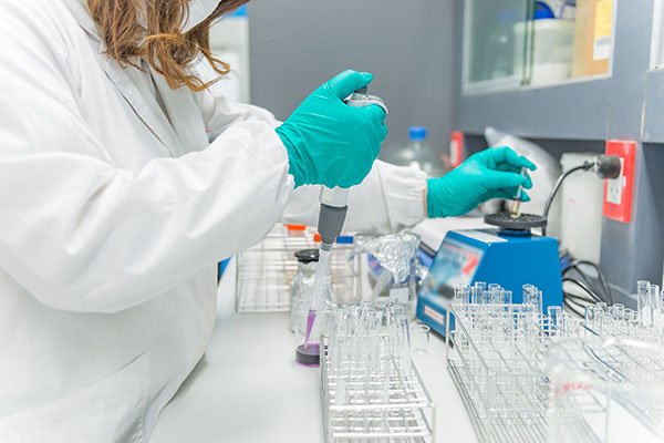
BOC Sciences is committed to providing customers with high-quality cell and organelle stains .
Cell staining and tracing reagents are important tools for tracking cells in complex environment. They are widely used in the study of cell migration, wound healing and stem cell differentiation. The research of cell movement and localization requires special stains, which must be non-toxic to living cells, and have a variety of fluorescent colors to choose at the same time, so as to match with different instrument laser wavelengths and filters, and can be co stained with antibody labeling or other cell analysis labeling probes.
Characteristics of Cell and Organelle Stains
Cell and organelle stains should have excellent core structure, stable reaction groups and good photostability to ensure the high stability and high labeling efficiency of biological coupling. It is used to monitor cell movement, localization, proliferation, migration, chemotaxis and invasion. This class of stains can be well retained in living cells for several generations and can display fluorescence for at least one week. The stains will transfer to daughter cells, but not to adjacent cells in the population. It is best to apply to both flow cytometry and fluorescence microscope.
Application of Cell and Organelle Stains
In the field of biomedical research, it is often necessary to selectively and real-time visualize biological small molecules, neutral molecules, metal ions, anions and organelles such as lysosomes, mitochondria and endoplasmic reticulum through cell imaging. Therefore, it is necessary to develop a variety of fluorescent probes and stains to meet different analysis and research needs.
- Probes targeting mitochondrial ROS have been widely used to study many disease-related mitochondrial activities. Due to the unique mitochondrial structure and double-layer membrane with negative membrane potential, the probe for mitochondria needs a positively charged scaffold, which should also be highly hydrophobic.
- Many studies have focused on monitoring the oxidation and cleavage function of lysosomes by measuring the pH and redox active chemicals of lysosomes. Lysosomal tracking dyes include a pH sensitive core that produces a fluorescent signal when protonated.
- The nucleus is the container of most DNA in the cell. Some conventional DNA application dyes are mainly DNA intercalators, which are used for the detection and quantification of nucleic acids.
- The most widely used part of targeting endoplasmic reticulum (ER) and golgi is phenylsulfonamide group, which can selectively bind to the rich cyclooxygenase (COX) in ER membrane. Therefore, this class of probes have similar scaffolds, phenylsulfonamide groups conjugated to fluorophores, can detect chemicals in ER and golgi matrix.
- Previously available probes targeting cell membranes generally share a common method, that is, combining environmentally sensitive fluorophores to generate membrane specific signals, and combining membrane fixed parts to minimize the diffusion of probes.
Resources

- Hoechst Dyes: Definition, Structure, Mechanism and Applications
- Mastering the Spectrum: A Comprehensive Guide to Cy3 and Cy5 Dyes
- Fluorescent Probes: Definition, Structure, Types and Application
- Fluorescent Dyes: Definition, Mechanism, Types and Application
- Coumarin Dyes: Definition, Structure, Benefits, Synthesis and Uses
- Unlocking the Power of Fluorescence Imaging: A Comprehensive Guide
- Cell Imaging: Definitions, Systems, Protocols, Dyes, and Applications
- Lipid Staining: Definition, Principles, Methods, Dyes, and Uses
- Flow Cytometry: Definition, Principles, Protocols, Dyes, and Uses
- Nucleic Acid Staining: Definition, Principles, Dyes, Procedures, and Uses
Online Inquiry
