

Hoechst Stain Dyes
Hoechst stains are part of a family of blue fluorescent dyes used to stain DNA. These Bis-benzimides were originally developed by Hoechst AG and numbered all compounds, so the dye Hoechst 33342 is the 33342th compound produced by the company. There are 3 related Hoechst dyes: Hoechst 33258, Hoechst 33342 and Hoechst 34580. The dyes Hoechst 33258 and Hoechst 33342 are the most commonly used dyes and they have similar excitation emission spectra.
 Figure 1. Chemical structure of Hoechst dyes.
Excitation emission spectrum of Hoechst dye.
Both dyes are excited by ultraviolet light at about 350 nm and both emit blue-green fluorescence with a maximum emission spectrum of 461 nm. Unbound dyes have maximum fluorescence emission in the 510-540 nm range. Hoechst stains can be excited with xenon or mercury arc lamps or UV lasers. The considerable Stokes shift between the excitation and emission spectra makes Hoechst dyes useful for experiments using multiple fluorophores. The fluorescence intensity of Hoechst dye also increases with the pH of the solvent. Hoechst dyes are soluble in water and organic solvents such as dimethylformamide or dimethylsulfoxide. The concentration can reach 10 mg/mL. In the dark, the aqueous solution is stable for at least six months at 2–6 ° C. For long-term storage, the solution is frozen at -20 ° C or lower.
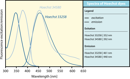 Figure 2. Excitation-emission spectra of Hoechst dyes.
The dye binds to the small groove of double-stranded DNA and preferentially selects sequences that are rich in adenine and thymine. Although dyes can bind to all nucleic acids, AT-rich double-stranded DNA strands can significantly enhance fluorescence. Hoechst dyes are cell-permeable and can bind to DNA in living or fixed cells. Therefore, these stains are often referred to as ultrafiltration membranes, meaning that living cells can survive the treatment with these compounds. Cells expressing specific ATP-binding cassette transporters can also actively transport these stains out of the cytoplasm.
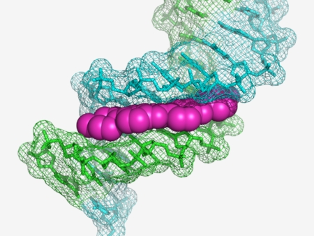 Figure 3. Hoechst 33258 (magenta) bound to the minor groove of DNA (green and blue).
Applications
DNA in bacteria or eukaryotic cells is usually stained using concentrations of 0.1–12 μg/ml. Cells were stained for 1-30 minutes at room temperature or 37 ° C and then washed to remove unbound dye. Green fluorescence of unbound Hoechst dye may be observed on samples stained or partially washed with too much dye. Hearst dyes are often used as a replacement for another nucleic acid dye called DAPI.
The main differences between Hoechst dye and DAPI are:
Hoechst dye is less toxic than DAPI, ensuring higher viability of stained cells.
The extra ethyl groups of Hoechst dyes make them more cell-permeable.
Nuclei staining dyes can make the cells viable after staining.
 Figure 4. Transmission image of HeLa cells, with overlay of Hoechst 33258 staining (blue).
Reference:
- Latt, SA.; et al. Recent developments in the detection of deoxyribonucleic acid synthesis by 33258 Hoechst fluorescence. Journal of Histochemistry and Cytochemistry. 1975, 23 (7): 493–505.
Resources

- Hoechst Dyes: Definition, Structure, Mechanism and Applications
- Mastering the Spectrum: A Comprehensive Guide to Cy3 and Cy5 Dyes
- Fluorescent Probes: Definition, Structure, Types and Application
- Fluorescent Dyes: Definition, Mechanism, Types and Application
- Coumarin Dyes: Definition, Structure, Benefits, Synthesis and Uses
- Unlocking the Power of Fluorescence Imaging: A Comprehensive Guide
- Cell Imaging: Definitions, Systems, Protocols, Dyes, and Applications
- Lipid Staining: Definition, Principles, Methods, Dyes, and Uses
- Flow Cytometry: Definition, Principles, Protocols, Dyes, and Uses
- Nucleic Acid Staining: Definition, Principles, Dyes, Procedures, and Uses
Online Inquiry
