
Green fluorescent protein Dyes
Green fluorescent protein (GFP) is a protein consisting of about 238 amino acids, which can be excited from blue light to ultraviolet light to emit green fluorescent light. Although many other marine organisms have similar green fluorescent proteins, traditionally, green fluorescent protein (GFP) refers to a protein that was first isolated from Victorian multitubular luminescent jellyfish. This protein was first discovered by Shimomura and others in Victoria's multitubular luminous jellyfish in 1962. This light-emitting process also needs the help of the cold-light protein jellyfish, and this cold-light protein can interact with calcium ions.
 Figure 1. Structure of the Aequorea victoria green fluorescent protein.
Introductions
The wild-type green fluorescent protein found in Victoria's multi-tube luminescent jellyfish, 395nm and 475nm are the maximum and second-largest excitation wavelengths, respectively, and the peak point of its emission wavelength is 509nm, which is greenish bluish in visible spectrum position. The fluorescence quantum yield (QY) of green fluorescent protein was 0.79. The green fluorescent protein obtained from sea pansy has a higher excitation peak only at 498 nm. In cell biology and molecular biology, the green fluorescent protein (GFP) gene is often used as a reporter gene. The green fluorescent protein gene can also be cloned into vertebrates (such as rabbits) for expression, and used to prove a hypothetical experimental method. Through genetic engineering technology, the green fluorescent protein (GFP) gene can be transferred into the genomes of different species and continuously expressed in the offspring. Now, the green fluorescent protein (GFP) gene has been introduced and expressed in many species, including bacteria, yeast and other fungi, fish (such as zebrafish), plants, flies, and even human mammalian cells.
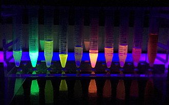 Figure 2. Different proteins produce different fluorescent colors when exposed to ultraviolet light.
Structure
Fluorescence is the most special feature of fluorescent proteins, and the chromophore plays a major role. The amino acids 65, 66, and 67 on the α-helix-serine, tyrosine, and glycine form chromophores after cyclization and dehydrogenation. Interestingly, the chromophore formation process is catalyzed by residues on the peripheral fence, and the substrate only requires oxygen. This implies the potential of green fluorescent protein to be widely used in different species: it can be independently expressed as a functional protein in different species without the need for additional factors. However, the exact process is still being discussed.The conjugated π bond on the chromophore can absorb the energy of the excitation light and release the energy with a longer wavelength of emitted light after a short time to form fluorescence.
Reference:
- Ormö M.; et al. Crystal structure of the Aequorea victoria green fluorescent protein. Science. 1996, 273 (5280): 1392–5.
Resources

- Hoechst Dyes: Definition, Structure, Mechanism and Applications
- Mastering the Spectrum: A Comprehensive Guide to Cy3 and Cy5 Dyes
- Fluorescent Probes: Definition, Structure, Types and Application
- Fluorescent Dyes: Definition, Mechanism, Types and Application
- Coumarin Dyes: Definition, Structure, Benefits, Synthesis and Uses
- Unlocking the Power of Fluorescence Imaging: A Comprehensive Guide
- Cell Imaging: Definitions, Systems, Protocols, Dyes, and Applications
- Lipid Staining: Definition, Principles, Methods, Dyes, and Uses
- Flow Cytometry: Definition, Principles, Protocols, Dyes, and Uses
- Nucleic Acid Staining: Definition, Principles, Dyes, Procedures, and Uses
Online Inquiry
